Adductor Canal: Difference between revisions
From WikiLectures
(image) |
m (Bot: Cosmetic changes) |
||
| (4 intermediate revisions by 2 users not shown) | |||
| Line 1: | Line 1: | ||
[[ | {{Dictionary | ||
|eng=Adductor canal, Hunter's canal | |||
|lat=Canalis adductorius | |||
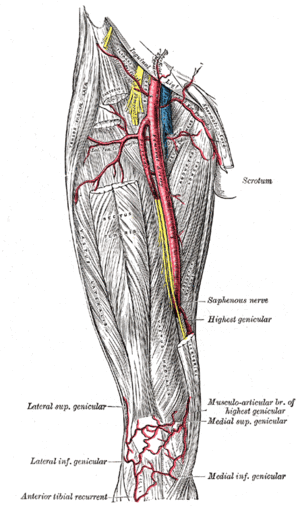
}}[[File:Gray550.gif|thumb|Adductor canal]] | |||
The '''adductor canal''' ('''Hunter‘s canal''', lat. ''canalis adductorius'') is bounded: | The '''adductor canal''' ('''Hunter‘s canal''', lat. ''canalis adductorius'') is bounded: | ||
* in front and laterally by [[ | * in front and laterally by vastus medialis (part of [[quadriceps femoris]]) | ||
* behind by [[adductor longus]] and [[adductor magnus]] | * behind by [[adductor longus]] and [[adductor magnus]] | ||
* covered in by a strong aponeurosis (lamina vastoadductoria) which extends from vastus medialis, across the femoral vessels to adductor longus and magnus | * covered in by a strong aponeurosis (lamina vastoadductoria) which extends from vastus medialis, across the femoral vessels to adductor longus and magnus | ||
| Line 10: | Line 13: | ||
* the [[saphenous nerve]] | * the [[saphenous nerve]] | ||
<noinclude> | |||
=== | == Links == | ||
* [[Adductor | === Related articles === | ||
* [[Adductor Hiatus]] | |||
=== | === Bibliography === | ||
* | * {{Cite | ||
| type = book | |||
| surname1 = Petrovicky | |||
| name1 = Pavel | |||
| others = yes | |||
| title = Anatomie s topografií a klinickými aplikacemi | |||
| subtitle = Sv. 1, Pohybové ústrojí | |||
| edition = 1 | |||
| location = Martin | |||
| publisher = Osveta | |||
| year = 2001 | |||
| range = 463 | |||
| isbn = 80-8063-046-1 | |||
}} | |||
</noinclude> | |||
[[Category:Anatomy]] | [[Category:Anatomy]] | ||
Latest revision as of 18:02, 8 December 2014
| English: Adductor canal, Hunter's canal Latin: Canalis adductorius |
The adductor canal (Hunter‘s canal, lat. canalis adductorius) is bounded:
- in front and laterally by vastus medialis (part of quadriceps femoris)
- behind by adductor longus and adductor magnus
- covered in by a strong aponeurosis (lamina vastoadductoria) which extends from vastus medialis, across the femoral vessels to adductor longus and magnus
The canal contains:
- the femoral artery and femoral vein
- the saphenous nerve
Links[edit | edit source]
Related articles[edit | edit source]
Bibliography[edit | edit source]
- PETROVICKY, Pavel, et al. Anatomie s topografií a klinickými aplikacemi : Sv. 1, Pohybové ústrojí. 1. edition. Martin : Osveta, 2001. 463 pp. ISBN 80-8063-046-1.


