Subdural hematoma: Difference between revisions
No edit summary |
(Translation from czech to english) |
||
| Line 1: | Line 1: | ||
__noTOC__ | __noTOC__ | ||
[[Image:Obaly mozku.png|400px|right|Mozkové pleny]] | [[Image:Obaly mozku.png|400px|right|Mozkové pleny]] | ||
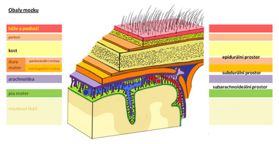
A subdural hematoma is a collection of blood between the [[Dura Mater|dura mater]] and the [[arachnoid]]. | |||
* '''''[[ | * '''''[[Acute subdural hematoma|Acute]]''''' – manifestation within 24−48 h after the injury, | ||
* ''''' | * '''''subacute''''' – manifestation within 3 weeks after the injury, | ||
* '''''[[ | * '''''[[Chronic subdural hematoma|chronic]]''''' – manifestations over weeks or months.<ref name="Ambler">{{Cite|isbn=80-7262-433-4|name1=Zdeněk|location=Prague|surname1=Ambler|year=2006|range=171-181|title=Základy neurologie|type=book|publisher=Galen|edition=6}}</ref> | ||
| | |||
| | |||
| | |||
| | |||
| | |||
| | |||
| | |||
| | |||
| | |||
| | |||
}}</ref> | |||
== | == Acute subdural hematoma == | ||
It is most often caused by bleeding of [[bridging veins]] (they lead from the surface of the brain to the sinus dura matris). It is almost always accompanied by [[cerebral contusion]] . | |||
=== | === Clinical picture === | ||
* | * As with [[epidural hematoma]] , but a more gradual course, | ||
* | * if a [[lucid interval]] is present , it tends to be longer, | ||
* [[ | * [[hemiparesis]] (usually on the opposite side to the hematoma, but can be on the same side if the brainstem has shifted), | ||
* [[ | * [[anisocoria]] - less often | ||
=== | === Diagnosis === | ||
* [[CT]]− | * [[CT]]− semilunar hyperdense in the [[calf]]. | ||
<gallery> | <gallery> | ||
Image:SD hematom dvoudobý 1.jpg|CT subdurální hematom dvoudobý, vpravo | Image:SD hematom dvoudobý 1.jpg|CT subdurální hematom dvoudobý, vpravo | ||
| Line 43: | Line 24: | ||
</gallery> | </gallery> | ||
=== | === Therapy === | ||
* | * Conservative (spontaneous resorption of the hematoma), | ||
* | * possibly neurosurgical treatment. | ||
== | == Chronic subdural hematoma == | ||
A so-called ''chronic subdural hematoma occurs'' when the blood hematoma becomes large . It has its own capsule and inside is a serous fluid . A chronic hematoma enlarges due to an osmotic mechanism (the capsule is a semipermeable membrane) and due to repeated minor subdural bleeding from proliferating capillaries on the hematoma membrane. It manifests clinically only after a long delay, for example in 3 months or 3 years.<ref name="Nevšímalová">{{Cite|isbn=80-7262-160-2|name1=Soňa|name2=Evžen|name3=Jiří|location=Prague|surname1=Nevšímalová|surname2=Růžička|surname3=Tichý|year=2005|range=163-170|title=Neurologie|type=book|publisher=Galen|edition=1}}</ref> | |||
| | Predisposing factors for the development of chronic subdural bleeding: older age, [[alcoholism]] , [[arachnoid cysts]] , [[coagulopathy]] , [[anticoagulant treatment]] , [[arterial hypertension]] , [[epilepsy]] ,...<ref name="Nevšímalová" /> | ||
| | |||
| | |||
| | |||
| | |||
| | |||
| | |||
| | |||
| | |||
| | |||
| | |||
| | |||
| | |||
}}</ref> | |||
=== | === Clinical picture === | ||
* [[ | * [[Headaches/PGS|Headaches]] , [[vomiting]] , eye congestion, | ||
* | * personality changes and deterioration of intellect, | ||
* | * quantitative [[Disorders of consciousness (pediatrics)|disorders of consciousness]] with hemiparesis. | ||
=== | === Prognosis === | ||
* | * The prognosis of a subdural hematoma depends on the severity of the head injury, the speed of treatment and the age of the patient. | ||
About 50% of people with large acute hematomas survive, although the injury often results in permanent brain damage. Younger people have a better chance of survival than the elderly. People with chronic subdural hematomas usually have the best prognosis, especially if they have few or no symptoms and have remained conscious and alert after a head injury. In the elderly, there is an increased risk of further bleeding (hemorrhage) after a subdural hematoma, because the elderly brain cannot expand again and fill the space where the blood was (a so-called subdural hygroma will then form in this space), so it is more vulnerable to future bleeding into the brain even when minor head injuries. [https://cs.medlicker.com/2018-subduralni-hematom#prognoza MUDr. Michal Vilímovský] | |||
<noinclude> | <noinclude> | ||
== | == Links == | ||
=== | === related links === | ||
* [[ | * [[Craniocerebral trauma]] | ||
* [[ | * [[Acute subdural hematoma]] | ||
* [[ | * [[Chronic subdural hematoma]] | ||
* [[ | * [[Epidural hematoma]] | ||
* [[ | * [[Subarachnoid hemorrhage]] | ||
=== Reference === | === Reference === | ||
<references/> | <references/> | ||
=== | === References === | ||
*{{ | *{{Cite|isbn=80-7262-160-2|name1=Soňa|name2=Evžen|name3=Jiří|location=Prague|surname1=Nevšímalová|surname2=Růžička|surname3=Tichý|year=2005|range=163-170|title=Neurologie|type=book|publisher=Galen|edition=1}} | ||
| | *{{Cite|isbn=80-7262-433-4|name1=Zdeněk|location=Prague|surname1=Ambler|year=2006|range=171-181|title=Základy neurologie|type=book|publisher=Galen|edition=6}} | ||
| | |||
| | |||
| | |||
| | |||
| | |||
| | |||
| | |||
| | |||
| | |||
| | |||
| | |||
| | |||
}} | |||
*{{ | |||
| | |||
| | |||
| | |||
| | |||
| | |||
| | |||
| | |||
| | |||
| | |||
| | |||
}} | |||
</noinclude> | </noinclude> | ||
Revision as of 00:31, 21 December 2022
A subdural hematoma is a collection of blood between the dura mater and the arachnoid.
- Acute – manifestation within 24−48 h after the injury,
- subacute – manifestation within 3 weeks after the injury,
- chronic – manifestations over weeks or months.[1]
Acute subdural hematoma
It is most often caused by bleeding of bridging veins (they lead from the surface of the brain to the sinus dura matris). It is almost always accompanied by cerebral contusion .
Clinical picture
- As with epidural hematoma , but a more gradual course,
- if a lucid interval is present , it tends to be longer,
- hemiparesis (usually on the opposite side to the hematoma, but can be on the same side if the brainstem has shifted),
- anisocoria - less often
Diagnosis
- SD hematom dvoudobý 1.jpg
CT subdurální hematom dvoudobý, vpravo
- SD hematom dvoudobý 2.jpg
CT subdurální hematom dvoudobý, vpravo
- SD hematom dvoudobý 3.jpg
CT subdurální hematom dvoudobý, vpravo
Therapy
- Conservative (spontaneous resorption of the hematoma),
- possibly neurosurgical treatment.
Chronic subdural hematoma
A so-called chronic subdural hematoma occurs when the blood hematoma becomes large . It has its own capsule and inside is a serous fluid . A chronic hematoma enlarges due to an osmotic mechanism (the capsule is a semipermeable membrane) and due to repeated minor subdural bleeding from proliferating capillaries on the hematoma membrane. It manifests clinically only after a long delay, for example in 3 months or 3 years.[2] Predisposing factors for the development of chronic subdural bleeding: older age, alcoholism , arachnoid cysts , coagulopathy , anticoagulant treatment , arterial hypertension , epilepsy ,...[2]
Clinical picture
- Headaches , vomiting , eye congestion,
- personality changes and deterioration of intellect,
- quantitative disorders of consciousness with hemiparesis.
Prognosis
- The prognosis of a subdural hematoma depends on the severity of the head injury, the speed of treatment and the age of the patient.
About 50% of people with large acute hematomas survive, although the injury often results in permanent brain damage. Younger people have a better chance of survival than the elderly. People with chronic subdural hematomas usually have the best prognosis, especially if they have few or no symptoms and have remained conscious and alert after a head injury. In the elderly, there is an increased risk of further bleeding (hemorrhage) after a subdural hematoma, because the elderly brain cannot expand again and fill the space where the blood was (a so-called subdural hygroma will then form in this space), so it is more vulnerable to future bleeding into the brain even when minor head injuries. MUDr. Michal Vilímovský
Links
- Craniocerebral trauma
- Acute subdural hematoma
- Chronic subdural hematoma
- Epidural hematoma
- Subarachnoid hemorrhage
Reference
References
- NEVŠÍMALOVÁ, Soňa – RŮŽIČKA, Evžen – TICHÝ, Jiří. Neurologie. 1. edition. Prague : Galen, 2005. 163-170 pp. ISBN 80-7262-160-2.
- AMBLER, Zdeněk. Základy neurologie. 6. edition. Prague : Galen, 2006. 171-181 pp. ISBN 80-7262-433-4.
Kategorie:Neurologie Kategorie:Chirurgie Kategorie:Neurochirurgie Kategorie:Patologie


