Occipital bone: Difference between revisions
No edit summary |
No edit summary |
||
| Line 61: | Line 61: | ||
| range= 516 | | range= 516 | ||
| page = 136-139 | | page = 136-139 | ||
}}CIHÁK, Radomír. ''Anatomy I. '' 2nd ed. Prague: Grada, 2001. 516 pp. pp. 136-139. | }}CIHÁK, Radomír. ''Anatomy I. '' 2nd ed. Prague: Grada, 2001. 516 pp. pp. 136-139. [[ISBN 978-80-7169-970-5]] . | ||
{{Navbox - kosti}} | {{Navbox - kosti}} | ||
</noinclude> | </noinclude> | ||
[[Kategorie:Anatomie]] | [[Kategorie:Anatomie]] | ||
Revision as of 18:55, 1 June 2023
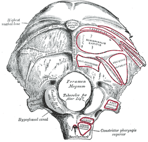
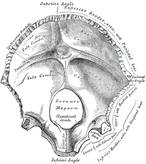
The occipital bone is the bone forming the back ( dorsal ) wall of the braincase. It resembles the shape of a triangular ladle, while being divided into several anatomical parts. In the middle of the base is the occipital opening, the foramen magnum , which ensures the passage of the medulla oblongata (and then the spinal cord itself ) and blood vessels to the spine .
Anatomical structures
Pars basilaris
The base is formed by the bone running ventrally from the foramen magnum to the sphenoid bone . Contains formations:
- clivus – a slope in the interior of the skull that rises from the occipital opening to the sphenoid bone, the a. vertebralis is placed on it ;
- tuberculum pharyngeal – a bump on the base of the skull where the pharynx attaches .
Partes laterales
The lateral parts of this bone are placed in pairs on the outside of the occipital foramen. They contain:
- condyli occipitales – a pair of elevations on which the atlas adjoins in the atlanto-occipital (joint of the carrier and occipital bone) joint ;
- canalis nervi hypoglossi – transversely through the condyles (transversely) the XII. hypoglossal cranial nerve ;
- fossa condylaris − fossa behind the condyle, sounds leads to the interior of the skull canalis condylaris;
- canalis condylaris – a channel containing the connections of extracranial and intracranial veins, located more dorsally behind the condyles;
- incisura jugularis – notch on the outer edge of the lateral parts, contains the processus jugularis, together with the adjacent temporal bone forms the jugular foramen ;
- processus jugularis − a projection from the pars lateralis behind the incisura jugularis, corresponds to the processus transversi vertebra;
- processus intrajugularis − divides the jugular foramen into ventromedial and dorsolateral parts.
Occipital scales
The scale of the occipital bone contains sections differentiated according to origin, i.e. a desmogenously ossified part and a chondrogenically ossified part. In essence, it can be said that the chondrogenic part is adjacent to the foramen magnum and the desmogenous part is the triangular remnant of the scale departing from the chondrogenic part.
The outer side of the scale contains:
- protuberantia occipitalis externa – a prominent bump (well palpable on the back of the head, especially in men), bony lines diverge from it:
- linea nuchalis suprema − the highest of the lines, the place of attachment of the superficial cervical fascia;
- linea nuchalis superior - close to the previous bar, for the insertion of the trapezius muscle ;
- linea nuchalis inferior – the most caudal, the place of attachment of the fibrous septa between the layers of the neck muscles.
- external occipital crest - for úchyt lig. bride
The inner side of the scale contains:
- protuberantia occipitalis interna – a similar bump as on the outside in the middle of the inner scale;
- eminentia cruciformis – bone elevations resembling a cross (sagittal-transverse);
- on them we can find impressions of venous vessels - sulcus sinus sagittalis superioris (cranially from the protuberance), sulcus sinus transversi (bilaterally from the protuberance), sulcus sinus sigmoidei (continuation of the transverse impression towards the temporal bone).
The most common variations
They arise mainly in the lambda seam and in the desmogenous part of the bone scale. These are ossa suturarum (Wormiana), i.e. separate bones in the seam. The desmogenous part may develop into a separate os interparietale. In the chondrogenic part, the paramastoideus process appears on the pars lateralis, a supernumerary vertebra around the foramen magnum or the atlas merging with the occipital bone.
Connection of the occipital bone with the surroundings
Ventrally, the occipital bone is connected by cartilage to the sphenoid bone in synchondrosis sphenooccipitalis , the connection changes to synostosis in the 18th–20th. year of life.
Sutura lambdoidea , or a saw-shaped seam in the shape of the letter λ , fastens the occipital bone with the parietal bone on the scale.
Links
Related articles
References
- CIHÁK, Radomír. Anatomy I. 2nd ed. Prague: Grada, 2001. 516 pp. pp. 136-139. ISBN 978-80-7169-970-5 .
Bones bones of the skull bones of the neurocranium os occipitale • os sphenoidale • os ethmoidale • os temporale • os frontale • os parietale • os lacrimale • os nasale • vomer bones of the splanchnocranium maxilla • os palatinum • os zygomaticum • mandible • os hyoideum • ossicula auditus • concha nasalis inferior axial skeleton spine • vertebrae • ribs • sternum • os sacrum bones of the upper limb plait scapula • clavicle arm and forearm humerus • ulna • radius hand carpus • metacarpus • finger bones bones of the lower limb plait os coxae ( hip bone • ischium • pubic bone ) thigh and lower leg femur • patella • tibia • fibula leg ossa tarsi • ossa metatarsi • bones of the fingers Portal: Anatomy


