Spinal cord: Difference between revisions
No edit summary |
No edit summary |
||
| Line 1: | Line 1: | ||
<!---------------------------------------------------------------------------------------------------------------- | {{DISPLAYTITLE:Spinal cord - structure of gray and white matter, cross section (draw scheme)}}<!---------------------------------------------------------------------------------------------------------------- | ||
* VLOŽENÝ ČLÁNEK | * VLOŽENÝ ČLÁNEK | ||
* Pozor – tento článek je využíván dalšími články, do kterých je vložen. Buďte prosím opatrní při jeho editaci: | * Pozor – tento článek je využíván dalšími články, do kterých je vložen. Buďte prosím opatrní při jeho editaci: | ||
| Line 10: | Line 10: | ||
* | * | ||
* Toto upozornění se vkládá šablonou {{subst:Vložený článek}} | * Toto upozornění se vkládá šablonou {{subst:Vložený článek}} | ||
-------------------------------------------------------------------------------------------------------------->The spinal cord is the center for simple reflexes. It runs through the spinal canal at the level of C<sub>1</sub>–L<sub>2</sub> (length 40–50 cm) and is covered by [[Spinal cord sheaths]]. It preforms '''reflex and conduction functions'''. The anterior and posterior spinal roots emerge from it, which subsequently join into a nerve bundle. The spinal cord contains '''mixed fibers''' (motor and sensitive) and '''vegitative fibers.''' | --------------------------------------------------------------------------------------------------------------> | ||
The spinal cord is the center for simple reflexes. It runs through the spinal canal at the level of C<sub>1</sub>–L<sub>2</sub> (length 40–50 cm) and is covered by [[Spinal cord sheaths]]. It preforms '''reflex and conduction functions'''. The anterior and posterior spinal roots emerge from it, which subsequently join into a nerve bundle. The spinal cord contains '''mixed fibers''' (motor and sensitive) and '''vegitative fibers.''' | |||
===Anatomy of the spinal cord=== | ===Anatomy of the spinal cord=== | ||
Revision as of 03:09, 25 December 2024
The spinal cord is the center for simple reflexes. It runs through the spinal canal at the level of C1–L2 (length 40–50 cm) and is covered by Spinal cord sheaths. It preforms reflex and conduction functions. The anterior and posterior spinal roots emerge from it, which subsequently join into a nerve bundle. The spinal cord contains mixed fibers (motor and sensitive) and vegitative fibers.
Anatomy of the spinal cord
At the height of C 2 –T 2 there is a thickening – intumescentia cervicalis and at the height of T 12 –L 1 there is intumescentia lumbosacralis (lumbalis) .
The spinal cord is divided into spinal segments . A spinal segment is a section of the spinal cord from which 1 pair of spinal nerves converge (a total of 31 pairs of spinal nerves – 8 cervical, 12 thoracic, 5 lumbar, 5 sacral, 1 coccygeal). It narrows caudally into a cone-shaped conus medullaris , the tip of which reaches L 1 –L 2 and then continues as a bundle of nerves, which we call the cauda equina.
Structure of the spinal cord
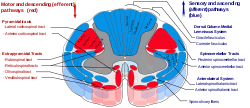
- White matter
-
- Funiculus lateralis, anterior and posterior – cords in which nerve fibers run upwards and downwards.
- Fissura mediana anterior – anterior notch between the anterior horns.
- Sulcus medianus posterior (posterior notch).
- Sulcus anterolateralis – the fibers of the anterior spinal roots emerge from it.
- Sulcus posterolateralis – exit of the posterior spinal root fibers.
- The spinal ganglion is located on the posterior spinal root (it surrounds the bodies of sensory neurons).
- Structure of the spinal cord
- Arranged in an H shape, formed by a cluster of neurons, it forms the anterior and posterior horns of the spinal cord . The lateral horns of the spinal cord form the columns columnae anteriores, laterales and posteriores (the anterior ones contain motoneurons, the lateral vegetative neurons, the posterior connecting neurons). The spinal canal – canalis centralis – runs through the center
Spinal reflexes
Spinal reflexes can occur at the level of the spinal cord. The spinal cord ensures the emptying of the bladder and rectum. The anatomical basis of the reflex is the reflex arc . The spinal cord is subordinate to the brain in its activity, it is the lowest reflex center in terms of development, its interruption means a loss of reflexes.
5 parts
- receptor, sensor;
- centripetal path (sensitive);
- CNS;
- centrifugal path (motor);
- effector.
spinal roots
The root fibers, fila radicularia ventralia , emerge from the sulcus ventrolateralis and merge into the anterior spinal roots – radices anteriores . From the dorsal part of the spinal cord, the fila radicularia dorsalia emerge and merge into the posterior spinal roots – radices dorsales . The roots enter the foramen intervertebrale and merge into the nervus spinalis .
The anterior spinal roots carry efferent fibers, the posterior spinal roots carry centripetal fibers. In terms of function, the anterior spinal roots are motor and the posterior spinal roots are sensitive.
Spinal nerves
- Cervical plexus (C 1 –C 4 ) – sensitively innervates the scalp and supraclavicular area, motorly innervates the neck muscles (e.g. n. phrenicus ).
- Brachial plexus (C 4 –Th 1 ) – innervation of the upper limb (e.g. radial nerve , ulnar nerve ).
- Thoracic nerves – pass within the intercostal spaces, do not form any plexuses. They innervate the chest wall.
- Lumbar plexus (L 1 –L 5 ) – innervation of the skin and muscles of the abdomen, thighs and pelvis.
- Sacral plexus (S 1 –S 5 ) – innervates the back of the thigh, buttocks, lower leg and foot. This plexus includes the thickest nerve in the human body, the sciatic nerve .
For more detailed information, see the Cervical plexus page
For more detailed information, see the brachial plexus page
For more detailed information, see the Thoracic plexus page
For more detailed information, see the Lumbar Plexus page
For more detailed information, see the Sacral Plexus page
Links
Související články
Externí odkazy
Použitá literatura
ČIHÁK, Radomír. ANATOMIE 3. second, revised and supplemented edition. Prague : Grada, 2004. 692 pp. ISBN 80-247-1132-X.


