Transmitted light microscopy
=== Transmitted light microscopy ===
- Indented line
Transmitted light microscopy is one of the techniques of the light microscopy. It's associated for any type of microscopy where the light is transmitted from a source on the opposite side of the specimen to the lens. This method it’s used to distinguish the morphologic characteristics and optics proprieties of the observed material.
- Indented line
The light rays are organized and precisely guided through the instrument that means that the light can be polarize and redirected.
- Indented line
The light from the condenser (has as function concentration of the light on the specimen) will fill the focal plane of the objective and will project a cone of light to illuminate the field of view, basically the condenser aperture diaphragm is responsible for controlling the angle of the illumination which permits the right balance of resolution and contrast in the microscope.
- Indented line
After the light passes through the specimen it goes to the objective where the image will be magnify and then it goes to the oculars when we can observe that enlargement. This type of microscopy permits the capture of high-quality images but the most important factor in this type of microscopy it’s the illumination in order to obtain a image bright enough to be useful.
2. Techniques requiring a transmitted light path:
2.1 Bright-field
- Indented line
It's the most usual method of microscopy, it helps to observe tissues because it makes the object appear against a bright background without glare and minimum heating of the specimenthe (it happens because the condenser is used to focus parallel rays of light on the specimen), this is caused by absorbance of some of the transmitted light in dense areas of the sample. the man who invented it Köhler, so it’s called Köhler illumination.
2.2 Dark-field
- Indented line
This method its use for biological samples as bacteria and micro-organisms, in this case the entrance of the light it’s bigger witch permit the diffraction of the lights rays and it’s illuminated obliquely. As a result, the field around the specimen is generally dark so the bright parts can be clearly observed.
2.3 Phase contrast
- Indented line
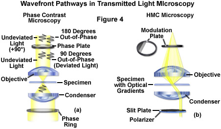
The light that is passing is slightly retarded so the specimen is illuminated by a hollow cone of light. It’s important in biology because reveals many cellular structures.
2.4 Polarisation
- Indented line
It uses polarised light to analyse structures that are birefringent (with two different indices of refraction). The image contrast came from the interaction between the plane-polarized light and the birefringent specimen. Polarised light microscopy as the function of measuring the amount of retardation that occurs in each direction and give information about the molecular structure of the birefringent object
2.5 Differential interference contrast optics
- Indented line
In this form of polarised light microscopy plane polarised beams of light are used to create a 3D-like image with shades of grey, a complex lighting scheme produces an image with the object appearing black to white on a grey background. Wollaston prisms situated in the condenser and above the objective produce the effect, and additional elements add colour to the image.

[1]


