Congenital hypertrophic pyloric stenosis


Hypertrophic pylor stenosis is an acquired diffuse hypertrophy and hyperplasia of the smooth muscle of the pylor and of the entire stomach.
Etiology
Ethiology is unknown, presumed participation of polygenic inheritance and environment, in 15% is proven familial occurrence. It is sometimes associated with hiatus hernia, esophageal atresia or Turner syndrome. The incidence of the disease varies greatly geographically, with around 1:5000 live births in the Czech Republic, up to five times more often in first-born boys. Affected are newborns and infants between 3-6 weeks of life, children older than 3 months rarely.
Clinical picture
- Explosive arc vomiting dominates, projectile (up to 1 m);
- Vomit contains acidic gastric juices, the content is digested milk, vomit is free from bile;
- there is dehydration, the child has a great appetite, he/she drinks eagerly; he/she loses weight (dehydration, insufficient caloric intake)
- the child is lethargic, constipated or hungry stools, has an age-like appearance;
- can lead to severe hypochloraemic alkalosis and hypokalaemia - severe condition, superficial breathing, loss of consciousness, convulsions - coma pyloricum;
- A peristaltic wave (left to right epigastrium) can be observed on the tummy immediately after drinking;
- with gentle palpation, about 70% of children have palpable resistance = olive, cherry size in epigastrium, right of midline - tumor pylori;
- icterus may occur rarely
Laboratory examination
- typically hypochloraemic alkalosis with hypokalaemia, hyponatraemia and dehydration;
- hypochloraemia can reach extreme values (below 75 mmol / l), its degree better reflecting potassium loss than potassium;
- elevated gastrin levels
Diagnostics
- clinical picture
- abdominal ultrasound
- the length (17 mm and more) and width (4 mm and more) of the pyloric channel are measured
- senzitivity 97%
- X-ray contrast examination (GIT passage) is currently used for diagnostic doubts
- stomach dilatation
- elongated and narrow pyloric canal (so-called rail or shoelace image)
- contrast medium must be aspirated with a nasogastric tube after examination (possible aspiration)
- rapid passage of contrast through the stomach eliminates pylorostenosis
- this test may reveal other causes of vomiting without bile:
- gastric atony
- delayed gastric emptying,
- gastroesophageal reflux
Differential diagnosis
- other causes of explosive vomiting:
- intracranial hypertension,
- pyloric atresia,
- antral membrane,
- stomach duplications,
- gastric atony
- delayed gastric emptying,
- gastroesophageal reflux;
- other causes of similar metabolic breakdown:
- acute adrenal insufficiency - bleeding or congenital hyperplasia - MAc,
- hyperkalemia,
- sodium loss in urine;
- hereditary metabolic disorders – Metabolic alkalosis can cause disorders of AMK metabolism (urea cycle disorder).
Therapy
- a conservative approach is not recommended
- the treatment is surgical
- longitudinal pyloromyotomy of hypertrophic pyloric muscle is most often performed (Weber-Ramstedt operation):
- it begins with a transverse laparotomy in the right part of the epigastrium
- the straight abdominal muscles and the oblique abdominal muscles are intersected longitudinally
- after opening the peritoneal cavity, a hypertrophic pylorus is luxated into the surgical wound
- a sharp longitudinal incision is made of serose and superficial muscle fibers (the incision starts 1-2 mm from the pyloroduodenal junction and ends in the area of the pyloral junction in the stomach)
- the muscle fibers are separated (along the entire length of the incision) by blunt dissection
- complication is perforation of the mucosa - this must be sutured with absorbable material and covered with omentum
- patients with more than 5% weight loss, metabolic alkalosis and hypochloraemia must parenterally rehydrate and correct the internal environment within 24 hours before surgery
- longitudinal pyloromyotomy of hypertrophic pyloric muscle is most often performed (Weber-Ramstedt operation):
- can also be performed by laparoscopic technique
- prognosis - good with timely operation.

Pyloromyotomy scar (rather large) 30 hrs post-op in a 1 month-old baby
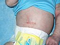
Horizontal cut 10 days after surgery (Psyloric Stenosis) in a 4 weeks old baby.


References
Related articles
- Oesophageal atresia
- Anal and rectal atresia
- Malrotation of volvulus and intestines
- Congenital Megacolon
Source
- BENEŠ, Jiří. Studijní materiály [online]. ©2007. [cit. 2010-04]. <http://www.jirben.wz.cz/>.
Literature
- HRODEK, Otto a Jan VAVŘINEC, et al. Pediatrie. 1. vydání. Praha : Galén, 2002. ISBN 80-7262-178-5.
- ŠAŠINKA, Miroslav, Tibor ŠAGÁT a László KOVÁCS, et al. Pediatria. 2. vydání. Bratislava : Herba, 2007. ISBN 978-80-89171-49-1.
- ŠNAJDAUF, Jiří a Richard ŠKÁBA. Dětská chirurgie. 1. vydání. Praha : Galén, 2005. ISBN 807262329X.
