Orthopantomography
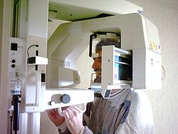
Orthopantomography is a radiodiagnostic method using RTG rays. Its result is an orthopantomogram (OPG) – a picture showing both jaws, dentition, jaw joints, jaw cavities and nasal cavity. This method is mainly used in dentistry. It belongs to extraoral imaging methods.
Imaging Technique
The technique is based on the combined rotational and translational movement of the X-ray tube and film and the screening of the rays by a vertical slit diaphragm.[1]
- Exposure approx. 15 s.
- Voltage: 55-85 kV.
- Current: 2-30mA.
During imaging, the patient's head is fixed in a cephalostat with the mouth slightly ajar. The X-ray and the film move in sync around the patient's head so that the resulting image is orthoradial.
Ortopantomogram
X-ray conditions
Head position
In order to obtain a high-quality image, the patient's head must be fixed in the cephalostat under the following conditions:
- The Frankfurt horizontal is respected,
- Head position is centered,
- The front teeth are in the focus zone of the device.
Today's devices have a light system that "navigates" to the correct fixation of the head.
Benefits of OPG
- Simplicity,
- An overview of dentition, jawbones and joints in a single image,
- 90% less surface radiation dose[1] compared to X-ray status (10 intraoral images),
- Option to compare right and left sides.
Disadvantages of OPG
- Shading of the frontal sections of the jaw of the spine – today the sensitivity in this section is automatically increased to improve the quality,
- Hypermetric image - magnification factor is 1.2-1.35.[2]
Links
Related Articles
References
- ↑ Jump up to: a b {{#switch: book |book = Incomplete publication citation. and George ZEMEN. Basics of radiodiagnostics and other imaging methods in dentistry. Prague : Karolinum, 1999. 978-80-7262-438-6. |collection = Incomplete citation of contribution in proceedings. and George ZEMEN. Basics of radiodiagnostics and other imaging methods in dentistry. Prague : Karolinum, 1999. {{ #if: 80-246-0005-6 |978-80-7262-438-6} } |article = Incomplete article citation. and George ZEMEN. 1999, year 1999, |web = Incomplete site citation. and George ZEMEN. Karolinum, ©1999. |cd = Incomplete carrier citation. and George ZEMEN. Karolinum, ©1999. |db = Incomplete database citation. Karolinum, ©1999. |corporate_literature = and George ZEMEN. Basics of radiodiagnostics and other imaging methods in dentistry. Prague : Karolinum, 1999. 978-80-7262-438-6} }
- ↑ Cite error: Invalid
<ref>tag; no text was provided for refs namedMushroom


