Disseminated intravascular coagulation
(Redirected from DIC)
Disseminated intravascular coagulation is a pathologically acquired state in which the vascular system is affected by upregulated coagulative activity that begins by the creation of tremendous amount of micro-thrombi in the capillary system, which leads to depletion of plasmatic coagulation factors, resulting in haemorrhage. DIC can be acute or chronic.
DIC is a secondary process as a result of tissue impairment.
Risk factors of disseminated intravascular coagulation:[edit | edit source]
- Sepsis
- Trauma (neural damage mostly)
- Tumor
- Severe post-transfusion reaction
- Rheumatism
- Gynecology and Obstetrics: amniotic fluid embolism, abruption of placenta, HELLP syndrome, eclampsia, dead fetus syndrome
- Severe liver failure
- Microcirculatory disorders (shock)
- Exposure of circulating blood to foreign materials (extracorporeal circulation) [1]
Organs that do mostly contribute to DIC[edit | edit source]
Due to high amounts of thromobokinase
4 P
- Pulmonary system (lungs)
- Prostate
- Pancreas
- Placenta [2]
In these pathological states molecules or cells with potential to cause DIC can enter the blood flow: Foreign tissue cells (aspiration of amnionic fluid in birth, severe trauma, operation, metastatic cells), pathological myeloid or lymphoid proliferative cells, endothelial cells and monocytes that are activated by cytokines (IL-1 and TNF) or endotoxin (in septic shock caused by G- bacteria), or cytoplasmic tissue factor from ruptured erythrocytes.
Pathophysiology[edit | edit source]
Coagulation of blood is physiologically a local process, whereas in DIC the coagulation is spread uncontrollably (as the name suggests) around the vascular system.
Main factors of DIC:
- Increase in thrombin production
- Anticoagulant mechanisms supression
- Fibrinolysis disorder
- Activation of inflammatory response
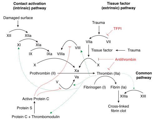
The whole process is started by activating the "external (extravascular) pathway" of coagulation - by activating plasma coagulation factor VII by binding to tissue factor III. Tissue factor is a molecule contained in the phospholipid membrane of cells and does not normally occur in the circulation. However, it is present on the surface of non-vascular cells and in the cytoplasm of some blood cells (blood cells do not express it on their surface). In addition to activating factor IX (activation of the "internal pathway" by components of the the external) and X, factor VII is also able to activate itself, promoting the whole reaction. Activation of factor IX further leads to increased production of activated factor X.
Activated factor X then leads to the conversion of prothrombin to thrombin and the subsequent cleavage of fibrinogen to fibrin monomer, which forms fibers (fibrin polymer) and leads to the formation of an intravascular "fibrin network". Thrombin also activates platelets. Activation of platelets includes changes in platelet shape, increased movement, release of granule contents, and aggregation [3]. This disseminated coagulation activity causes micro-embolization to the periphery, thereby significantly impairing organ perfusion and aiding in the development of ischemia in the affected areas.
The coagulation and anticoagulation system is in balance under normal circumstances. Coagulation is regulated by negative feedback between the individual stages of the coagulation cascade and by circulating coagulation inhibitors. The most effective inhibitor is antithrombin III, which inhibits their activity by binding to thrombin and other factors of the coagulation cascade (IXa, Xa, XIa, XIIa). The effect of antithrombin III represents about 3/4 of the antithrombotic activity. The remaining 25% are factors such as α2-macroglobulin, heparin cofactor II and α1-antitrypsin[3]. The activity of antithrombin III is increased by the presence of acidic proteoglycans such as heparin. Antithrombin-bound heparin alters its conformation and allows binding to more substrates. Thrombin further binds to thrombomodulin and converts protein C to active protein C, which in combination with its cofactor protein S, degrades activated coagulation factors V and VIII [3].
The formed fibrin chains are cleaved by the activated plasminogen - plasmin, which is activated by the action of the tissue plasminogen activator tPA. Cleaved fibrin chains can be detected in blood as fibrin degradation products (FDPs), also known as fibrin split products, which are blood components produced by clot degeneration.[4] The most notable subtype of fibrin degradation products is D-dimer. Fibrin and fibrinogen degradation product (FDP) testing is commonly used to diagnose disseminated intravascular coagulation.[5] In DIC, the processes of coagulation and fibrinolysis are dysregulated, and the result is widespread clotting with resultant bleeding.
Coagulation inhibitors are consumed in the process of DIC. Decreased inhibitor levels will permit more clotting so that a positive feedback loop develops in which increased clotting leads to more clotting. At the same time, thrombocytopenia occurs and this has been attributed to the entrapment and consumption of platelets. Clotting factors are consumed in the development of multiple clots, which contributes to the bleeding seen with DIC.
Simultaneously, excess circulating thrombin assists in the conversion of plasminogen to plasmin, resulting in fibrinolysis. The breakdown of clots results in an excess of FDPs, which have powerful anticoagulant properties, contributing to hemorrhage. The excess plasmin also activates the complement and kinin systems. Activation of these systems leads to many of the clinical symptoms that patients experiencing DIC exhibit, such as shock, hypotension, and increased vascular permeability. The acute form of DIC is considered an extreme expression of the intravascular coagulation process with a complete breakdown of the normal homeostatic boundaries. DIC is associated with a poor prognosis and a high mortality rate.
Coagulation and anticoagulation processes are closely related to the inflammatory response, and many proteins involved in the coagulation chain are also proteins of the acute phase of the inflammatory response. In the development of DIC, both coagulation and anticoagulant activity takes place, but also an inflammatory reaction that further deepens DIC.
Antithrombin is consumed during DIC to inhibit coagulation and it is also cleaved by enzymes produced by neutrophils activated by the inflammatory response. In addition, antithrombin production in the liver may be impaired as a result of liver damage due to insufficient perfusion and ischemia caused by micro-embolizations in the hepatic vessels [6]. Anticoagulant activity is also impaired by the consumption of other coagulation and anticoagulation factors. Inflammatory cytokines reduce thrombomodulin expression on cell membranes. Therefore, fibrinolysis and anticoagulation cannot keep pace with increasing coagulation activity, leading to further micro-embolization into tissues, development of ischemia, organ damage, development of inflammation and SIRS and MODS and finally depletion of coagulation and anticoagulant factors which leads to subsequent bleeding with shock. Inflammatory activity supports this by increasing permeability of blood vessel walls and following leakage of fluids from the intravascular space.
Course of DIC[edit | edit source]
Acute form[edit | edit source]
Acute form of DIC can be induced by infection, sepsis, severe trauma, burns, hemolytic transfusion reaction or liver failure and more. Its onset is measured in minutes to hours. Either bleeding or thrombotic process may predominate (depending on the nature of the underlying disease and the initial state of coagulation), the bleeding can be mild (bleeding from punctures) or dramatic, life-threatening - as severe as purpura fulminans - extensive skin bleeding associated with fever and hypotension. The microcirculatory thrombosis affects mostly kidneys, liver, lungs and central nervous system and can add up to a multiorgan failure. Gangrene may develop in the periphery because of embolic occlusion of large vessels of the limbs. [7]
Acute DIC can be divided into four consecutive phases:
- Initial stage (trigger stadium) - The beginning of activation of the coagulation pathway underlined by the stated risk factors. In this stadium theres hyper-coagulation, but so far without changes in the laboratory markers.
- Compensated DIC (hypercoagulation phase) - Incipient changes in laboratory tests, incipient fibrinolysis and increased consumption of coagulation factors
- Manifested subacute DIC (hyper-fibrinolytic phase) - Decreased coagulation, haemorrhagic diathesis, increased consumption of coagulation factors and increased fibrinolysis, typical changes in laboratory tests
- Decompensated DIC - Massively reduced coagulation, hemorrhagic diathesis, massive fibrinolysis, typical changes in laboratory tests. [8]
Chronic form[edit | edit source]
Chronic DIC can be based upon cancer, extensive vascular malformations or autoimmune diseases, its duration is in days or weeks. In most cases there are few to no symptoms, but can be diagnosed by performing blood coagulation exam. Under certain circumstances may become acute. [7]
Symptoms[edit | edit source]
The combination of defect in coagulation (reduced concentration of procoagulant factors) with a defect of primary hemostasis (thrombocytopenia) leads to disorders in the following systems:
Circulation: spontaneous and uncontrollable bleeding, petechiae and subcutaneous bleeding, diffusely localized thrombosis.
Cardiovascular system: hypotension, tachycardia, development of shock.
Nervous system: neurological symptoms depending on locus of lesion (consequences of micro-embolization), consciousness disorder.
GIT: melena, hematemesis.
Urogenital system: hematuria, metrorrhagia, oliguria. [9]
This condition can be complicated by embolizations into various organ systems - mainly kidneys (acute renal failure), lungs (ARDS) and brain vessels (stroke) are at risk. Another complication is development of systemic shock.
Diagnosis[edit | edit source]
Diagnosis of DIC involves a combination of laboratory tests and clinical evaluation. Laboratory findings suggestive of DIC include a low platelet count, elevated D-dimer concentration, decreased fibrinogen concentration, and prolongation of clotting times such as prothrombin time (PT). [10]
Laboratory markers:
- aPTT and PT coagulation is prolonged resp. increased
- The plasmatic levels of antithrombin III and factors V and VII are reduced
- Increased concentration of FDPs (fibrin degradation products, FDP) and D-dimers (the specificity of these is limited by the fact that both markers are increased in conditions such as trauma, surgery or thromboembolism)
- Decreased fibrinogen concentration
- Thrombocytopenia [11]
None of these are specific markers so a scoring range may help with diagnosis:
| Risk factors | Sepsis, trauma, Gynecology and Obstetric complications associated with DIC |
| Lab results | Thrombocytes level, FDPs, fibrinogen, AT III, aPTT, PT |
| Scoring |
Thrombocytes > 100 0 points, < 100 1 point, < 50 2 points Fibrin degradation products (FDP) no change 0 points, slightly elevated 2 points, massively elevated 3 points Elongated PT < 3 s 0 points, 3–6 s 1 point, > 6 s 2 points Fibrinogen > 1 g/l 0 points, < 1 g/l 1 point |
Scoring 5 and more with positive risk factors does incline toward DIC. Scoring should be repeated daily for patients in the high-risk group.[11]
Differentially diagnostic alternatives include other consumptive coagulopathy, trauma, major blood loss, surgery and subsequent replacement of volume together with dilution of coagulation factors, thrombocytopenia, hemolytic uremic syndrome (HUS), idiopathic thrombocytopenic purpura or heparin-induced thrombocytopenia [11].
Prognosis[edit | edit source]
Prognosis varies depending on the underlying disorder, and the extent of the intravascular thrombosis (clotting). The prognosis for those with DIC, regardless of cause, is often grim: between 20% and 50% of patients will die.[12] DIC with sepsis (infection) has a significantly higher rate of death than DIC associated with trauma.[10]
Therapy[edit | edit source]
The therapy of is complicated due to two opposing processes ongoing (clot formation vs depletion of coagulation factors and haemorrhage) and should be based upon which of these prevails. Even with top notch medical support is the morbidity in patients with ongoing DIC sadly high. Basic therapeutic approaches listed:
- Therapy of underlying disease - elimination of the cause of DIC.
- Circulation stabilization, adequate ventilation support, ensuring diuresis.
- Treatment of coagulation disorder - interruption of coagulation pathway (antithrombin, heparin - in chronic cases) and supplementation of missing blood components in order to achieve sufficient levels of coagulation factors, fibrinogen and platelets:
Platelet concentrate - to maintain the platelet count optimally > 50 × 109 per liter
Fresh frozen plasma - in case of bleeding and prolongation of PT
Fibrinogen - when below 1 g/l
Antithrombin - when chronic DIC, in order to reach 100-120% of antithrombin III levels
Heparin - disclaimed by some, used in the chronic form of DIC
Activated protein C
Recombinant activated factor VII. [7]
Fresh frozen plasma contains all pro- and anti-coagulation factors (concentration will vary from donor to donor). However, FFP also contains significant amounts of water, albumin and other plasma proteins, therefore transfusion of large amounts of plasma may cause decompensation, especially in patients with heart failure because of the rise in intravascular volume.
Some authors do consider the administration of heparin to be ineffective and dangerous - heparin must not be administered to patients which have developed haemorrhage because of DIC! For heparin to work, plasmatic concentration of AT III must be sufficient (at least 70% of default value). Therefore, it is necessary to know AT III levels and possibly supplement it. This goes as well for coagulation factor concentrates (PPSB), which could restart coagulation if the levels of AT III are inadequate.[8]
Links[edit | edit source]
Video[edit | edit source]
References[edit | edit source]
- ↑ LEVI, M, E DE JONGE a T MAYES. New treatment strategies for disseminated intravascular coagulation based on current understanding of the pathophysiology. Annals of Medicine. 2004, roč. 36, vol. 1, s. 41, ISSN 1365-2060.
- ↑ HEROLD, Gerd, et al. Herold Innere Medizin. 1. vydání. 2008. 895 s. s. 123-4. ISBN 3890197043.
- ↑ Jump up to: a b c MURRAY, Robert K, Daryl K GRANNER a Peter A MAYES, et al. The Harper´s Illustrated Biochemistry. 23. vydání. East Norwalk : Appelton & Lange, 1993. 872 s. s. 718. ISBN 80-7319-013-3.
- ↑ GAFFNEY PJ, EDGELL T, Creighton-Kempsford LJ, Wheeler S, Tarelli E (1995). "Fibrin degradation product (FnDP) assays: analysis of standardization issues and target antigens in plasma". Br. J. Haematol. 90 (1): 187–94. doi:10.1111/j.1365-2141.1995.tb03399.x. PMID 7786784.
- ↑ "Fibrin/Fibrinogen Degradation Products". <https://web.archive.org/web/20080821020612/http://peir.path.uab.edu/coag/article_77.shtml>. Archived from the original on 2008-08-21. Retrieved 2007-10-28.
- ↑ LEVI, M a E DE JONGE. Disseminated intravascular coagulation: What's new?. Critical Care Clinics. 2005, roč. 21, vol. 3, s. 67, ISSN 07490704.
- ↑ Jump up to: a b c LEBL, J, J JANDA a P POHUNEK, et al. Klinická pediatrie. 1. vydání. Galén, 2012. 698 s. s. 562-564. ISBN 978-80-7262-772-1.
- ↑ Jump up to: a b HECK, Michael a Michael FRESENIUS. Repetitorium Anästhesiologie. 5. vydání. Heidelberg : Springer, 2007. 642 s. ISBN 978-3-540-46575-1.
- ↑ BECKER, Joseph U. Disseminated Intravascular Coagulation [online]. Poslední revize 2009-09-10, [cit. 2010-05-13]. <https://emedicine.medscape.com/article/199627-overview>.
- ↑ Jump up to: a b LEVI M. Diagnosis and treatment of disseminated intravascular coagulation. Int J Lab Hematol. 2014;36(3):228-236. PubMed
- ↑ Jump up to: a b c BECKER, Joseph U a Charles R WIRA. Disseminated Intravascular Coagulation: Differential Diagnoses & Workup [online]. Poslední revize 10.9.2009, [cit. 2010-06-30]. <https://emedicine.medscape.com/article/199627-diagnosis>. Insert non-formatted text here.
- ↑ BECKER, Joseph U and Charles R Wira. Disseminated intravascular coagulation at eMedicine, 10 September 2009


