WikiLectures:Inferior vena cava - course and tributaries, cavocaval anastomoses
(Redirected from Inferior vena cava - course and tributaries, cavocaval anastomoses)
The inferior vena cava (IVC) is the largest vein of the human body. It is located at the posterior abdominal wall on the right side of the aorta. The IVC’s function is to carry the venous blood from the lower limbs and abdominopelvic region to the heart.
Course and Tributaries :
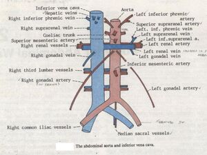
The inferior vena cava arises from the confluence of the common iliac veins at the level of L5 vertebra, just inferior to the bifurcation of the abdominal aorta. It then ascends the posterior abdominal wall, to the right side of the aorta and the bodies of the L3-L5 vertebrae. After passing through its fossa (caval fossa) on the posterior liver surface, the IVC enters the thorax by traversing the inferior vena caval foramen of the diaphragm.
The tributaries of the IVC correspond to the branches of the abdominal aorta. Note that some professors will want you to know at which vertebral level the IVC gets its direct tributaries, so they are as follows:
- The direct tributaries are the inferior phrenic veins (T8), right suprarenal (L1), renal (L1), right testicular (gonadal) (L2), lumbar (L1-L5), common iliac (L5) and hepatic (T8). If you want an easy way to remember them just memorize the mnemonic ' Portal System Returns To Liver In Humans'.
- Left gonadal and left suprarenal renal veins drain first into the left renal vein
- The veins of the stomach, spleen, pancreas, small and large intestines first empty into the hepatic portal vein. The hepatic portal vein carries this blood to the liver to be processed and detoxified. Then, the blood reaches the IVC through the hepatic veins.
The inferior vena cava communicates with the superior vena cava through the collateral vessels, which include the azygos vein, lumbar veins, and vertebral venous plexuses.
Hepatic Veins : Right, Middle and Left
Cavocaval Anastomoses :
Cavocaval anastomoses are venous connections between vena cava superior and vena cava inferior.
- v. cava superior – v. subclavian – v. internal thoracic – v. superior epigastric – v. inferior epigastric – v. external iliac – v. cava inferior
- v. cava superior – v. subclavian – vv. thoracoepigastric, v. lateral thoracic, vv. costoaxillary – v. superficial epigastric – v. great saphenous – v. femoral – v. external iliac – v. cava inferior
- v. cava superior – plexus venous vertebral – v. cava inferior
- vena cava superior – v. azygos and hemiazygos – vv. lumbales – v. cava inferior
These connections are particularly functional in restricting flow through the normal bloodstream. When obstructing the trunk, the veins thus provide an alternative flow path, most often in the area of the anterior abdominal wall.
- https://www.kenhub.com/en/library/anatomy/inferior-vena-cava
- PASTOR, Jan. Langenbeck's medical web page [online]. [cit. 11.3.2009]. <https://langenbeck.webs.com/>.

