Urinary Bladder
(Redirected from Urinary bladder)
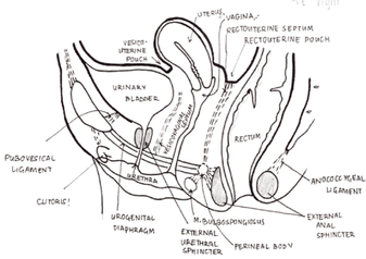
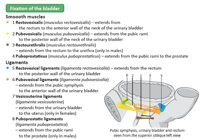
Urinary bladder - structure and position, fixation and syntopy in male and female (draw scheme)
Introduction[edit | edit source]
The urinary bladder is a pelvic organ that collects and holds urine before urination. It serves as a temporary reservoir for urine produced by the kidneys. When empty, it lies completely within the pelvic cavity, but enlarges upward into the abdominal cavity when full. It is the most anterior pelvic organ, located just behind the pubic bones and pubic symphysis.
The urinary bladder serves two primary functions: temporary storage of urine and assisting in its expulsion during micturition (urination). Structurally, it consists of three main parts: the body, fundus, and neck.
The body of the bladder, covered by peritoneum, collects urine, while the fundus is the bottom part, situated near the prostate and rectum in males, and close to the vagina in females. The neck of the bladder marks the convergence of the fundus and is continuous with the urethra. The apex of the bladder, directed towards the pubic symphysis, is connected to the umbilicus by the median umbilical ligament, a remnant of the urachus.
Syntopy[edit | edit source]
• Anterior – pubic symphysis
• Posterior – rectum in males, vagina in females
• Superior – small intestine in peritoneal cavity, uterus in females
• Inferior – prostate in males, urogenital diaphragm in females
-Urine enters the bladder via the right and left ureters, and internally these orifices are marked via the trigone (triangular area in fundus)
Trigone of bladder – triangular like area in the anteroposterior part of bladder. The mucosa here is not folded. In females trigone projects on anterior part wall of vagina.
Bladder musculature[edit | edit source]
The bladder wall contains specialized smooth muscle known as the detrusor muscle, consisting of inner plexiform, middle circular, and external longitudinal layers. The detrusor muscle is innervated by the parasympathetic system. The bladder also features the sphincter vesicae, a single layer of circular smooth muscles around the neck of the bladder, innervated by the sympathetic system, which prevents semen from flowing into the bladder during ejaculation.
Spaces, fasciae and septae[edit | edit source]
The spaces, fasciae, and septae surrounding the urinary bladder provide structural support and organization within the pelvic cavity.
The transversalis fascia is located anterior to the apex of the urinary bladder. It forms the retropubic space, which is the area between the bladder and the pubic symphysis.
The vesicoumbilical fascia of Delbert is a triangular-shaped fascia situated between the medial umbilical ligaments and the median umbilical ligament, extending to the umbilicus.
Covering the superior surface of the bladder is the parietal peritoneum. In males, this arrangement creates the rectovesical pouch, a space between the rectum and the bladder. In females, it forms the vesicouterine pouch, a space between the uterus and the bladder. Additionally, there are paravesical fossae, peritoneum-lined spaces on both sides of the bladder.
The paravesical space surrounds the urinary bladder within the pelvic cavity, located between the bladder and the pelvic walls.
Two septae are also present: the rectovesical septum, a plate of connective tissue between the bladder and the rectum, and the vesicovaginal septum, a plate of connective tissue between the bladder, vagina, and adjacent cervix of the uterus.
References[edit | edit source]
- HUDÁK, Radovan – KACHLÍK, David. Memorix anatomie. 2. edition. Triton, 2013. ISBN 978-80-7387-712-5.


