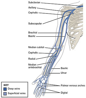
Superficial and Deep Veins of Extremities
Veins of upper limb[edit | edit source]
Superficial veins[edit | edit source]
The major superficial veins of the upper limb are the cephalic and basilic veins. They are located within the subcutaneous tissue of the upper limb.
Basilic Vein[edit | edit source]
The basilic vein originates from the dorsal venous network of the hand and ascends the medial aspect of the upper limb.
At the border of the teres major, the vein moves deep into the arm. Here, it combines with the brachial veins from the deep venous system to form the axillary vein.
Cephalic Vein[edit | edit source]
The cephalic vein also arises from the dorsal venous network of the hand. It ascends the antero-lateral aspect of the upper limb, passing anteriorly at the elbow.
At the shoulder, the cephalic vein travels between the deltoid and pectoralis major muscles (known as the deltopectoral groove), and enters the axilla region via the clavipectoral triangle. Within the axilla, the cephalic vein empties into axillary vein.
The cephalic and basilic veins are connected at the elbow by the median cubital vein.
Deep veins of upper limb[edit | edit source]
Deep venous system of upper limb lies underneath the deep fascia. It consists of paired veins (up until the axilla) following arteries of the same names. The biggest deep vein of upper limb is the brachial vein. Pulsations of the brachial artery assist in venous return. Veins like that are knows as vena comitantes. A network of perforating veins connects superficial and deep systems.
- Digital palmar veins
- radial veins
- ulnar veins
- brachial veins
Veins of lower limb[edit | edit source]
Superficial veins of lower limb[edit | edit source]
Superficial veins of lower limb run in the subcutaneous tissue. The two major superficial veins are great saphenous vein and small saphenous vein. dorsal venous arch of the foot is a venous plexus collecting blood from the back of the foot and the plantar region via the venous plantar network, the great saphenous vein is formed from the medial side of the plexus and the small saphenous vein is formed from the lateral side.
The great saphenous vein is formed by the dorsal venous arch of the foot, and the dorsal vein of the great toe. It ascends medially to the leg, passing anteriorly to the medial malleolus, and posteriorly to the medial condyle at the knee. It receives tributaries from small superficial veins of the leg and drains into the femoral vein inferiorly to the inguinal ligament. It can be used in coronary artery bypass surgery.
The small saphenous vein is formed by the dorsal venous arch of the foot, and the dorsal vein of the little toe. It moves up the posterior side of the leg, passing posteriorly to the lateral malleolus, along the lateral border of the achilles tendon. At the level of the knee, the small saphenous vein passes between the two heads of the gastrocnemius muscle and drains into the popliteal vein in the popliteal fossa.
Deep veins of lower limb[edit | edit source]
Deep veins of lower limb are located below the deep fascia. The deep veins run along the arteries of the same names and are often located in the same vascular sheath which allows for the pulsations of arteries to aid in venous return.
The anterior tibial vein forms from the penetrating branches of dorsal venous arch of the foot.
The posterior tibial vein forms from the medial and lateral plantar veins and accompanies the posterior tibial artery, entering the leg posteriorly to the medial malleolus.
On the posterior surface of the knee, the anterior tibial, posterior tibial and fibular veins form the popliteal vein which enters the thigh via the adductor canal. After that it is known as the femoral vein it lies anteriorly to the femoral artery. The femoral vein leaves the thigh running beneath the inguinal ligament and after passing it becomes the external iliac vein.
The deep vein of the thigh drains the thigh muscles like the quadriceps femoris and empties into the distal section of the femoral vein.
Used literature[edit | edit source]
- ČIHÁK, Radomír. Anatomie III. 2., upr. a dopl vydání. Praha : Grada Publishing, spol. s. r. o., 2004. 673 s. ISBN 80-247-1132-X.
- GRIM, Miloš a Rastislav DRUGA. Základy anatomie. 5., Anatomie krajin těla. 1. vydání. Praha : Galén : Karolinum, 2002. ISBN 80-7262-179-3.
- STANDRING, Susan, et al. Gray's Anatomy : The Anatomical Basis of Clinical Practice. 39. vydání. London : Elsevier Ltd, 2005. ISBN 0-443-07168-3.


