Action potential in the heart
Cardiomyocytes[edit | edit source]
Brief description[edit | edit source]
Cardiomyocytes form a type of striated muscle. Cardiac cells are grouped into functional syncytia, they are connected to each other by intercalary discs. Each cell is Y-shaped.
Electrical properties[edit | edit source]
The resting membrane potential is around −70 to −90 mV, i.e. negative inside compared to the surroundings. The stimulation produces an action potential, which is different from the action potential of the muscle. Depolarization develops rapidly, as in skeletal muscle, but there is a plateau phase before the potential returns to its original level.
Functions of ions[edit | edit source]
The extracellular concentration of K+ ions affects the resting membrane potential, while the extracellular concentration of Na+ ions affects the magnitude of the action potential.
Action potential course[edit | edit source]
- Action potential phase
- depolarization;
- plateau;
- repolarization.
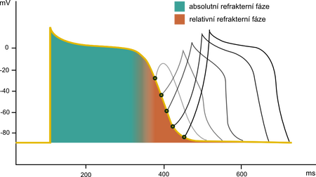
The rapid depolarization s caused by the opening of voltage-gated channels for Na + (phase 0 in the figure). Then there is a rapid repolarization zcaused by the closing of these channels (phase 1 in Fig.). The following plateau phase jis caused by the slower opening of Ca2+ channels, which are also controlled by voltage (phase 2 in Fig.). Thus, final repolarization to the resting potential j is possible only after the closure of these channels and also the efflux of K+ ions through different types of channels (phases 3 and 4 in Fig.). By a relatively long absolute refractory phase, when it is not possible to evoke an action potential even with a suprathreshold stimulus, the myocardium is protected from too high a frequency of contractions and also from changing the direction of propagation of the action potential back. In the relative refractory phase, irritation can only be induced by a suprathreshold stimulus (see figure).
Channels[edit | edit source]
Cardiomyocytes contain in the membrane 3 types of ion channels important for the formation of an action potential:
- fast Na+ channels;
- fast K+ channels;
- slow Na+/Ca2+ channels.
- Potassium channels
The first one accounts for the early outflow current. The second allows the entry of K+ during the plateau phase. At the same time, it prevents its exit, allowing it only at lower membrane potentials. The third type is slowly activated. The current builds up over time and causes depolarization.
Sodium is voltage controlled and has two gates. The outer ones open at a membrane potential of −70 to −80 mV. The inner ones then close, thereby inactivating the Na+ channel. The slow Ca2+ channel is then activated at a membrane voltage of around −30 až −40 mV.
The speed of action potential propagation[edit | edit source]
The conduction velocity of the impulse in the myocardium is 0,3–0,5 m/s (1/250 of the impulse velocity in large nerves and 1/10 of the velocity in skeletal muscle). The speed of propagation in the cells of the conduction system of the heart (Purkinje fibers) is higher, up to 4 m/s.
Pacemaker potentials[edit | edit source]
Brief description[edit | edit source]
We call the nodus sinuatrialis and nodus atrioventricularis a pacemaker, because they are able to create an action potential and thereby set the rhythm of the heart.
Action potential[edit | edit source]
Their resting voltage after each completed action voltage drops to the threshold value (beginning of phase 4 in Fig.), at which this prepotenciál (phase 4 in Fig.) triggers the next excitation. In this phase, the K+, current , which induced repolarization by opening its channels at the end of the action potential, weakens. At the same time, Ca2+ channels open. In the heart, they have 2 subtypes, the T channel (transient) and the L channel (long lasting). Current through T channels terminates diastolic depolarization, and current through L channels induces a new action voltage.
Action potential in the cells of the transmission system[edit | edit source]
The formation of an action potential in the cells of the conduction system has a different character than in the working myocardium. It is caused by the inactivation of fast Na + channels. At a membrane potential that is higher than −55 mV, the internal gate of sodium channels closes, thereby inactivating them. Since the resting potential in the cells of the conduction system is −55 mV, the sodium channels remain inactivated and do not fire an action potential. When the potential level reaches −40 mV, only slow Na + /Ca 2+ channels open to cause an action potential. After a few ms , potassium channels close and open, restoring the resting potential. In the working myocardium the onset of the action potential is fast and sharp, whereas in the conduction system, due to the inactivation of fast Na + channels, the onset of the action potential is slower and the magnitude of the depolarization is lower.
Odkazy[edit | edit source]
Související články[edit | edit source]
- Cardiac conduction system
- Pacemaker potential
- Action potential (physiology)
- Heart
- Resting membrane potential
- Electrocardiography
- Ion channels
- Cardiomyocyte
External links[edit | edit source]
- Action potential and ECG - Free ECG book
References[edit | edit source]
- GUYTON, Arthur C and John E. (John Edward) HALL. Textbook of medical physiology. 11th edition. Philadelphia : Elsevier Saunders, c2006. ISBN 0-7216-0240-1 .
- GANONG, William F. Ganong's review of medical physiology. 23rd edition. Boston, Mass: McGraw-Hill Medical, 2010. ISBN 978-007-127066-3 .
- GANONG, William F, et al. Review of Medical Physiology. 20th overall, 1st in Galen edition. Prague: Galén, 2005. 890 pp. ISBN 80-7262-311-7 .


