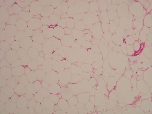
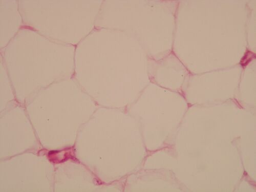
Adipose tissue (histology slide)
Overview[edit | edit source]
Preparation 1[edit | edit source]
Name: White adipose tissue HE
Description: White adipose tissue cells have a nucleus on the periphery. The cell contains one large fat drop, cytoplasm forms only a thin strip on the periphery. The cell has the appearance of a signet ring. The cells form lobules separated by dense collagenous tissue. Adipose tissueis richly vascularized and innervated.
Preparation 2[edit | edit source]
Name: White adipose tissue HE
Description: When processing a histological section, fat (triglycerides) is washed out of the cell, the central part of the cell is therefore empty. the lipid droplet is not separated from the cytoplasm by a membrane. Cytoplasm forms only a thin strip at the periphery. Fat cells are surrounded by the lamina basalis.
Connective tissue[edit | edit source]
Brown adipose tissue (preparation)
Adipose tissue (histology slide)
Articular cartilage (preparation)
Desmogenous ossification (preparation)
Enchondral ossification (preparation)


