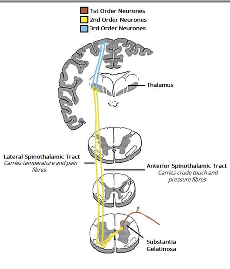
Anterolateral system of sensitive spinal tract
Contains Info about:
Anterolateral system of sensitive spinal tracts - (spinothalamic, spinoreticular and spinotectal tracts), pain pathways.
The Anterolateral System mediates primitive sensations, characterized by lower myelination and diverse sensory modalities, transmitting signals at lower velocities. Similar to the dorsal column-medial lemniscus pathway, the anterolateral spinothalamic tracts involve three groups of neurons:
- The first neuron originates in the dorsal root ganglia, with its peripheral process connecting to sensory receptor endings, while its central process enters the spinal cord through the posterior root to synapse with the second-order neuron.
- The second neuron's axon decussates and ascends to higher levels.
- The third neuron, typically located in the thalamus, projects a fiber to a sensory region of the cerebral cortex.
While this three-neuron chain is the most common arrangement, some afferent pathways may involve more or fewer neurons. Many neurons in these ascending pathways exhibit branching and provide significant input to the reticular formation, which in turn activates the cerebral cortex. The transmission of sensory information involves several pathways:
- Lateral Spinothalamic Tract: Responsible for conveying painful and thermal sensations as it ascends through the spinal cord.
- Anterior Spinothalamic Tract: Conveys crude touch and pressure sensations.
- Spinotectal Tract: Carries pain, thermal, and tactile information to the superior colliculus of the midbrain, facilitating spinovisual reflexes.
- Spinoreticular Tract: Provides a pathway from muscles, joints, and skin to the reticular formation.
- Spino-Olivary Tract: Offers an indirect route for afferent information to reach the cerebellum.
Antero-lateral Spinothalamic Tract[edit | edit source]
Pain impulses are transmitted to the spinal cord through two types of fibers: fast-conducting fibers (A-delta fibers) and slow-conducting fibers (C fibers). Fast-conducting fibers promptly alert individuals to initial sharp pain, while slow-conducting fibers are responsible for prolonged burning or aching pain.
The first-order neurons originate from sensory receptors in the periphery via A-delta fibers (fast-conducting) and C fibers (slow-conducting). They enter the spinal cord from the dorsal root ganglion, travel to the tip of the posterior gray column, ascend 1-2 levels, and terminate at the substantia gelatinosa in the dorsal horn, forming the posterolateral tract of Lissauer.
Second-order neurons convey sensory information from the substantia gelatinosa, decussating to the opposite side of the central nervous system. Upon decussation, they bifurcate into two fibers:
- Crude touch and pressure fibers enter the anterior spinothalamic tract.
- Pain and temperature fibers enter the lateral spinothalamic tract.
Although functionally distinct, these tracts run alongside each other and can be viewed as a single pathway - the spinal lemniscus. They travel in their respective pathways, synapsing in the thalamus.
Third-order neurons carry sensory signals from the thalamus to the primary sensory cortex of the brain. Originating from the ventral posterolateral nucleus of the thalamus, they ascend through the internal capsule, ultimately terminating at the sensory cortex.
As the spinothalamic tract ascends through the spinal cord, new fibers are added to its medial aspect. Consequently, in the upper cervical segments, sacral fibers are predominantly lateral, while cervical segments are mostly medial.
Ascending through the medulla oblongata, the anterior and lateral spinothalamic tracts accompany the spinotectal tract, collectively forming the spinal lemniscus.
Spinotectal Tract[edit | edit source]
The process begins with axons entering the spinal cord from the dorsal root ganglion, journeying towards the gray matter where they synapse with second-order neurons whose exact identity remains unspecified.
These second-order neuron axons cross the median plane and ascend as the spinotectal tract within the anterolateral white column, closely paralleling the path of the lateral spinothalamic tract. As they traverse through the medulla oblongata and pons, they culminate their journey by synapsing with neurons in the superior colliculus of the midbrain.
This pathway serves a crucial role in facilitating spinovisual reflexes, contributing to movements of the eyes and head towards the source of the stimulation.
Spinoreticular Tract[edit | edit source]
The axons enter the spinal cord from the dorsal root ganglion, terminate on unknown second-order neurons in the gray matter. The axons from these 2nd order neurons ascend the spinal cord as the spino-reticular tract in the lateral white column, mixed with the lateral spinothalamic tract. Most of the fibers are uncrossed and terminate by synapsing with neurons of the reticular formation in the medulla oblongata, pons, and midbrain.
The spinoreticular tract provides an afferent pathway for the reticular formation, which plays an important role in influencing levels of consciousness


