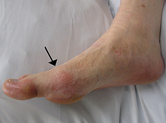
Arthritis uratica (gout)
Arthritis uratica (gout, podagra) is a joint disease caused by a disorder of purine metabolism. This leads to the accumulation of the final product of their degradation - uric acid - in the form of crystals and subsequently to inflammatory and degenerative changes in the joints. It affects men in 90% , most often in the 4th-5th decade, it affects women almost exclusively after menopause (loss of uricosuric effect of estrogens).
- Primary (idiopathic) gout
- represents 90% of cases.
- Secondary gout
- is a concomitant symptom of another disease (eg. leukemia, kidney disease, cell breakdown in cytostatic treatment, Lesch-Nyhan syndrome).
The form of gout can be acute or chronic.
Secondary hyperuricemia[edit | edit source]
With increased uric acid production or decreased excretion - hyperuricaemia, which is the cause of gout.
Metabolic hyperuricemia[edit | edit source]
It is caused by increased production of uric acid. The cause may be:
- Increased cell lysis
- leading to increased nucleic acid turnover. E.g. in leukemia, many more cells die than normal, and thus more purines are degraded, from which uric acid is formed as the end product.
- Increased purine intake
- mostly meat diet, offal, legumes, cocoa.
- Disorders of some enzymes
- eg. increased PRPP-synthase activity, hypoxanthine-guanine-phospho-ribosyl-transferase (HGPRT) deficiency - Lesch-Nyhan syndrome, glucose-6-phosphatase defect.
Renal hyperuricemia[edit | edit source]
Decreased urinary excretion of uric acid also leads to hyperuricaemia. We will meet him, for example, at:
- chronic renal failure;
- use of loop diuretics;
- increased levels of lactate, ketone bodies and some other anions - there is competition for a transporter that serves for tubular secretion of uric acid.
Pathogenesis of gout[edit | edit source]
The basic mechanism is the induction of an inflammatory reaction by the crystals. Uric acid crystallizes more easily in poorly perfused tissues, in colder parts of the body and in more acidic environments. Therefore, the tissues of the acral joints are mainly affected. During a gout attack, crystals fall from the supersaturated synovial fluid into the joint cavity and are subsequently coated with a protein (mostly IgG). The coated crystal then causes inflammation - the crystals are phagocytosed by neutrophils . Since they cannot be degraded, the phage cannot decompose them in their phagosome. The phagocyte breaks down and lysosomal enzymes are released. This deepens the tissue damage. In addition, the pH drops, making urates crystallize more easily. Nucleic acids are released from the disintegrating cells, which then form additional uric acid. In addition, activated macrophages release cytokines and inflammatory mediators that stimulate collagenase release by chondrocytes and synovialocytes
Clinical picture[edit | edit source]
We distinguish 4 clinical stages :
Asymptomatic hyperuricemia[edit | edit source]
- The patient has an elevated serum uric acid level, but there is no arthritis or gout, and the patient has no problems.
- It can last a lifetime
Acute gouty arthritis[edit | edit source]
- Recurrent seizures (most often at night from full health, often after exertion or dietary error), which do not leave significant changes on the joints after disappearance, but their repetition accumulates smaller changes and this condition is called chronic gout.
- The pathogenetic process probably proceeds in such a way that in hyperuricemia, uric acid crystallizes in the synovial membrane and fluid. These crystals are taken up by synovial cells, which have a phagocytic capacity. Subsequently, chemotactic (IL-1 and TNF-alpha) stimulates chondrocytes and other synovial cells to produce collagenase, which damages the articular cartilage. Neutrophilic granulocytes are also chemotactically induced - they break down after phagocytosis and thus release other lytic enzymes and free radicals, which destroy cartilage and cause inflammation in the joint.
- articular cartilage and synovium are permeated with sodium urate;
- the affected joint is swollen (most often metatarsophalangeal joint of the big toe - podagra), severely painful, the skin above it reddish and warm (ie all Celsus symptoms of inflammation), the joint cavity contains a small amount of serous exudate containing uric acid.
- Acute attack is accompanied by general symptoms of inflammation - fever , leukocytosis, increased FW, CRP.
- The attack usually disappears after a few days, within 2 weeks at the latest.
Intercritical period[edit | edit source]
Asymptomatic interval between seizures, usually 6-24 months.
Chronic gouty arthritis[edit | edit source]
In the joint, around it and in the epiphyses of the bones, nodular rigid deposits with urate crystals = gout tophae develops (especially the joints of the limbs, the auricle, olecranon, tendons, extensors of the small joints of the hand). Microscopically, these are deposits containing urate needles assembled into star-shaped formations with a giant cell inflammatory reaction of the foreign-body type.
Diagnostics[edit | edit source]
For certain detection, it is necessary to detect urate crystals in the synovial fluid, a tofu finding containing sodium urate deposits (chemical detection by a murexy test or in a polarizing microscope).
Laboratory marks
- Hyperuricaemia (in men> 416 μmol / l, in women> 360 μmol / l), inflammatory joint effusion, determination of uric acid in urine in 24 hours.
- X-ray - sharply demarcated bone erosion ("as punched out"), near the joints of an extensive osteolytic lesio
Complications[edit | edit source]
They are renal - the crystals settle in the canals and later pass into the interstitium, leading to hydronephrosis (pressure atrophy of the kidney from stagnant urine). An important secondary gout is Lesch-Nyhan syndrome in a hypoxanthine-guanine phosphoribosyltransferase defect, which affects men, damages the CNS (chorea, athetosis, self-numbing mental retardation, etc.). There is an increased incidence of gout in other diseases - arterial hypertension (60%), hypertriacylglycerolemia (80%), nephropathy (urolithiasis , interstitial gout nephritis, acute renal failure).
Therapy[edit | edit source]
Treatment of acute gout attack[edit | edit source]
They are currently recommended for the treatment of acute gout [1]
- glucocorticoids administered orally or parenterally,
- nonsteroidal antirheumatic drugs,
- colchicine
Colchicine[edit | edit source]
Colchicine is a traditional but still used treatment. If therapy is started within 24 hours of the onset of an attack, it is at least as effective as other[1]. A standard dry extract of Colchicum is administered. There are several dosing regimens. It usually starts with a loading dose of 1 mg, after which additional doses of 0.5 mg are given every 1 to 2 hours up to a total daily dose of 1.5 to 2 mg. In the following days, doses of 0.5-1 mg per day are continued as needed[2].
Glucocorticoids[edit | edit source]
Glucocorticoids can be used to treat gout, either orally (eg. prednisone 30-40 mg daily) or intra-articularly (eg. triamcinolone ) or parenterally (eg. methylprednisolone 20 mg i.v. 2x per day)[1]
Nonsteroidal antirheumatic drugs[edit | edit source]
Especially in younger patients without comorbidities and with a low risk of gastrointestinal side effects, the administration of nonsteroidal antirheumatic drugs is a reasonable alternative to glucocorticoid treatment. For example, indomethacin 50 mg 3x per day[1], ibuprofen or diclofenac are used .
Chronic therapy:[edit | edit source]
- dietary measures – reducing foods rich in purines (offal, seafood, legumes), reducing alcohol intake, reducing overweight;
- uricostatics – blockers of uric acid synthesis (xanthine oxidase blockers) - allopurinol, in case of its contraindications, intolerance or insufficient efficacy febuxostat.
- uricosurics – increase the excretion of uric acid by the kidneys - probenecid, benzbromarone
In case of nephrolithiasis or nephropathy, only uricostatics can be administered.
When a patient is treated with xanthine oxidase blockers, he must not take purine analogues such as azathioprine.
Aciduric renal infarction[edit | edit source]
It occurs during leukocyte breakdown (in newborns or after cytostatic leukemia therapy). Uric acid crystals are deposited in the renal tubules. Macroscopically - rusty brown stripes in papillae.
Pseudogout[edit | edit source]
Pseudogout (also arthritis calcinosa, chondrocalcinosis) - a designation for all conditions that are clinically reminiscent of gout (arthritis attacks), but in which calcium phosphate, calcium pyrophosphate, calcium hydroxyapatite are deposited in the joint and adjacent areas (it is a pathological calcification - von Kossa stain). I is possible to observe a giant cell type reaction from foreign bodies.
Links[edit | edit source]
Related articles[edit | edit source]
External links[edit | edit source]
Source[edit | edit source]
- PASTOR, Jan. Langenbeck's medical web page [online]. [cit. 2009]. <https://langenbeck.webs.com/>.
References[edit | edit source]
- KLENER, Pavel. Vnitřní lékařství. 4. edition. Praha : Galén, Karolinum, 2011. 1174 pp. ISBN 978-80-7262-705-9.
- ↑ Jump up to: a b c d BECKER, Michael A – GAFFO, Angelo L. UpToDate : Treatment of gout flares [online]. Wolters Kluwer, The last revision 2019-05-30, [cit. 2019-07-19]. <https://www.uptodate.com/contents/treatment-of-gout-flares>.
- ↑ Česká republika. Státní ústav pro kontrolu léčiv. Souhrn údajů o přípravku : Colchicum-Dispert 500 mikrogramů obalené tablety. 2017. sp.zn. sukls67270/2009 a sp.zn. sukls99384/2015, sukls170192/2015.


