Audiometry
| There are multiple articles on WikiScripts for this query. |
Audiometry is a diagnostic method that monitors auditory function disorders , with the help of which a doctor can determine hearing loss. According to the detected type of hearing impairment, it can partially determine its cause.
Subjective audiometry[edit | edit source]
The doctor is dependent on data from the patient, the method is based on how the patient perceives tones and subsequently signals.
Tone audiometry[edit | edit source]
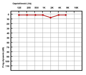
It is a diagnostic examination of the hearing aid, carried out using a tone generator at individual frequencies. It determines the hearing threshold for pure tones in the frequency range of 125 to 8000 Hz with the possibility of regulating the sound intensity level from -10 dB to 100 dB. This examination allows us to determine the sensitivity threshold of the human ear.
Course of audiometric examination[edit | edit source]
The patient is placed in a so-called quiet chamber, which is soundproofed from the surrounding noise. He has headphones or a bone vibrator on. The doctor sends tones to the patient, which he gradually intensifies - from a sound intensity of -10 dB, gradually adding 5 dB. When he hears the examined tone, he signals the doctor with a button. The doctor notes the threshold level of intensity for the given tone and continues the examination with another tone with a higher frequency. We first perform the examination on the healthy ear. If the examined person hears normally in both ears, we start with the right ear. We perform the test using both air and bone conduction.
Air ducts[edit | edit source]
The patient has headphones on. We successively test eight frequencies (1 kHz, 2 kHz, 3 kHz, 4 kHz, 6 kHz, 8 kHz, 500 Hz, 250 Hz). ear blue cross .
Bone conduction[edit | edit source]
The patient is fitted with a bone vibrator that vibrates the mastoid process. We successively test seven frequencies (1 kHz, 2 kHz, 3 kHz, 4 kHz, 6 kHz, 500 Hz, 250 Hz). The doctor marks the lowest signal level to which the patient responded as follows: for the right ear a red arrow <, for the left ear a blue arrow >.
Evaluation of the tone audiogram[edit | edit source]
Perceptual hearing disorder - symmetrical hearing loss in both bone and air conduction. There is usually more of a drop in the higher notes.
Conductive hearing loss - loss in air conduction, while bone conduction is normal. Mixed hearing loss - a combination of the two previous ones
Objective audiometry[edit | edit source]
Due to age or mental well-being, the doctor cannot rely on the data provided by the patient. The method is based directly on sensing biosignals.
Impedance audiometry = tympanometry[edit | edit source]
The essence of the method is to measure the amount of acoustic energy reflected from the eardrum. Part of the energy falling on the eardrum is transmitted further to the middle ear system and further to the inner ear. However, some of the acoustic energy is reflected from the eardrum. The measurement takes place by inserting a probe with three passing tubes into the external ear canal. The first tube is connected to a miniature earphone, from which a measurement signal with a frequency of 226 Hz and a sound pressure intensity of 85 dB is fed into the space of the external ear canal. The second tube is connected to a measuring microphone that detects the magnitude of the reflected measuring signal. The third tube causes a change in air pressure using a special air pump in the range of +200 daPa to -600 daPa from the atmospheric pressure value. The result of the measurement is a tympanogram.
Otoacoustic emissions (OAE)[edit | edit source]
This examination method is based on the ability of hair cells in the organ of Corti to produce a very faint sound in response to an acoustic stimulus, which can be picked up by a sensitive microphone. This sound is called an otoacoustic emission . These acoustic emissions are caused by short impulses. If the examined person has a hearing defect, there will be no response or a delayed response to otoacoustic emissions in the ear.
Investigation using evoked potentials[edit | edit source]
The method consists in measuring the bioelectric activity of the auditory pathway, which can be sensed on the surface of the head as an evoked auditory potential. These potentials arise in response to an acoustic stimulus. Using this method, we monitor bioelectrical impulses along the entire length of the pathway (in the cochlea, auditory nerve , brainstem and cerebral cortex ).
- ECoG - electrocochleography - examination of the evoked responses of the cochlea
- BERA – Brainstem Evoked Response Examination
- CERA – examination of evoked responses of the cerebral cortex
Sources[edit | edit source]
- ŠVECOVÁ, V.. The use of compensatory aids in the integration of students with hearing impairment [online]. ©2010. [cit. 2013-30-11]. <https://theses.cz/id/mn8zs3/82393-920274139.pdf>.
- STANICKÝ, O.. Audiometry [online]. ©2011. [cit. 2013-30-11]. <https://dspace.vutbr.cz/bitstream/handle/11012/20387/Audiometrie_Stanick%C3%BD_78003.pdf?sequence=2>.
- BLAHÁK, P.. Audiometer for pure tone audiometry [online]. ©2010. [cit. 2013-30-11]. <https://dspace.vutbr.cz/bitstream/handle/11012/15790/Audiometr_pro_audiometrii_cistymy_tony.pdf?sequence=-1>.
- SIEMENS AUDIOLOGICKÁ TECHNIKA S.R.O.,. Subjektivní audiometrie [online]. ©2010. [cit. 2013-30-11]. <http://hearing.siemens.com/cz/cs/children/testing-children/subjective-audiometry/subjective-audiometry.html>.
- VITALION,. Audiometry [online]. ©2010. [cit. 2013-30-11]. <https://vysetreni.vitalion.cz/audiometrie/>.
References[edit | edit source]
Amler E. et al. Practical tasks from biophysics I. Institute of Biophysics UK, 2nd Faculty of Medicine, Prague 2006

