Basal ganglia
Basal ganglia (nuclei basales) are part of the gray matter of the endbrain outside the thalamus, immersed in the white matter of the brain. These are developmentally old structures. They are used in the creation and control of movement, they are also involved in cognitive functions and the functions of the limbic system.
Morphology[edit | edit source]
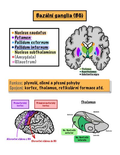
Morphologically, the basal ganglia primarily include large nuclei in the basal part of the telencephalon. They are[1]:
- caudate nucleus; - long, arched C-shaped, lateral to the lateral ventricle, broad head - caput, directed upwards as corpus and continues dorsolaterally as cauda
- putamen - lateral and basal from nc. caudatus
- globus pallidus – we divide it into:
- globus pallidus medialis (pallidum internum) – output basal ganglion;
- globus pallidus lateralis (pallidum externum) – vintrinsic basal ganglion.
The Nucleus caudatus together with the putamen is referred to as the corpus striatum and the putamen together with the globus pallidus forms the nucleus lentiformis.
The Striatum is divided into: striatum ventrale - rostral part, nc. accumbens + tuberculum olfactorium, important in the feeling of euphoria, satisfaction
striatum dorsale - larger part, the fibers coming from other parts to the basal nuclei end here
Pallidum is divided into: pallidum ventrale - smaller, front part, under the commissura anterior
pallidum dorsale - the larger part above the anterior commissure
The name is derived from the stripes ( striae ) of gray matter that connect these two nuclei through the white matter of the capsula interna.
Developmentally, thecorpus amygdaloideumalso belongs to the basal ganglia, which, however, from a functional point of view is more closely related to the limbic system.[1]
- Pathways of the basal nuclei
- Cortical circuit - they connect with the cortex of the telencephalon
- sensorimotor- one part of the fibers connects the basal nuclei bidirectionally with motor and sensitive ones
- association circle
- Limbic circuit
- Extracortical pathways - smlead to the brainstem to the structures from which the extrapyramidal pathways descend (tectum, nc. ruber, formatio reticularis..)
náhled|330x330pixelů|Bazální ganglia Next we describe[2]:
- substantia innominata Reicherti – a group of structures basal to the basal ganglia consisting of
- striatum ventrale / nucleus accumbens – ventral parts of the striatum;
- pallidum ventrale – ventral part of the globus pallidus;
- rostral (medial and central) nuclei of the corpus amygdaloideum;[1]
- nucleus basalis Meynerti – scattered gray matter formed by neurons producing acetylcholine;
- nucleus subthalamicus (corpus Luysi) – a nucleus located in the subthalamu basally from zona incerta, involved as a so-called intrinsic (intermediate) nucleus in the circuit of the basal ganglia;
- substantia nigra – a dark nucleus located in the mesencephalon. We distinguish:
- pars compacta – dopamine-producing monoaminergic nucleus for the corpus striatum (A9 in the system of monoaminergic nuclei);
- pars reticularis – output basal ganglion.
Function[edit | edit source]
The basal ganglia are involved in a circuit. The general scheme is: cortex → input basal ganglion → output basal ganglion → thalamus → cortex. Division of basal ganglia according to involvement [1][3]:
- basal ganglia input:
- they receive information from the cerebral cortex;
- their neurons are inhibitory ( GABA mediator );
- corpus striatum (ncl. caudatus, putamen, striatum ventrale = ncl. accumbens septum);
- output basal ganglia:
- they send information via the thalamus to the cerebral cortex or directly to the brainstem brainstem (reticular formation);
- their neurons are also inhibitory (GABA);
- globus pallidus medialis, pallidum ventrale (→ cortex) and substantia nigra, pars reticularis (→ trunk);
- intrinsic basal ganglia:
- they transfer information between input and output cores in the so-called indirect path;
- globus pallidus lateralis (inhibitory neurons – GABA);
- ncl. subthalamicus (excitatory neurons – glutamate);
- they modulate the activity of the corpus striatum and direct/indirect pathways via dopamine – pars compacta substantiae nigrae.
- they transfer information between input and output cores in the so-called indirect path;
Based on which cortical area the stimuli come to the basal ganglia circuit, which nuclei are used as input and output, and which functional cortical area the projection then goes to, we describe the four loops of the basal ganglia.
Loops of the basal ganglia[edit | edit source]
| Bark → | → Intup BG → | → Outup BG → | → Nuclei of the thalamus → | → Bark | Function | |
|---|---|---|---|---|---|---|
| Sensorimotor | primary motor (4), premotor and supplementary motor (6), somatosensitive (3, 1, 2) | putamen | globus pallidus medial, substantia nigra - part ret. | ncl. anterior ventrals (VA) (ncl. lateral ventrals (VL)) | supplementary motor (6) | basic motor patterns |
| Oculomotorics | frontal oculomotor field (8), prefrontal, secondary visual (18, 19) | ncl. caudatus | globus pallidus medial, substantia nigra - part ret. | ncl. anterior ventrals (VA) ncl. mediodorsals (MD) | frontal oculomotor field (8) | motor of the eyeball |
| Associational | prefrontal, posterior parietal, premotor (6) | head ncl tailed | globus pallidus medial, substantia nigra - part ret. | ncl. anterior ventrals (VA) ncl. mediodorsals (MD) | prefrontal (6) | spatial memory |
| Limbic | orbitofrontal (11, 12, 47), limbic cortical areas (hippocampal formation, gyrus cinguli) | ventral striatum (ncl. accumbens), head ncl. tailed | ventral pale | ncl. mediodorsales (MD) (ncl. ventral anteriors (VA)) | orbitofrontal (11, 12, 47), area cincularis ant. | motor response of the limbic system |
Direct and indirect track[edit | edit source]
The basal ganglia are involved in the circuit in the direct and indirect pathways. In the case of a sensorimotor loop, they consist of[[2]:
- direct path: cortex – striatum – globus pallidus medialis (pallidum internum) – thalamus – cortex;
- indirect track: cortex– striatum – globus pallidus lateralis (pallidum externum) – ncl. subthalamicus – globus pallidus medialis – thalamus – cortex.
The basal ganglia are involved in the control of motor functions and partly also in cognitive functions. They generally have a dampening effect on motor skills - on the one hand, through a direct feedback effect on the neurons of the motor cortex, and on the other hand, by forward damping of cortical stimuli through the reticular formation.
The role of the basal ganglia in motor control[edit | edit source]
The basal ganglia play an important role in motor control. They are activated before the start of the movement, so they probably participate in its planning. [4] Uhey are also said to be involved in the control of complex movement patterns, such as writing, playing with a ball or speaking.[5] Zapojení bazálních ganglií|thumb Information coming from primarily motor and somatosensitive areas of the cerebral cortex enters the putamen. They continue[2]:
- direct pathway: the putamen inhibits the globus pallidus medialis with GABA. Globus pallidus medialis is also inhibitory, with its high spontaneous activity dampens the thalamus (GABA). If it is itself inhibited, the inhibition of the thalamus is reduced and it acts excitatory (with the help of glutamate) on the cerebral cortex (mainly the supplementary motor area). Increased activity in this pathway therefore results in higher locomotor activity. Its function is therefore to support movements.
- indirect pathway: the putamen inhibits the globus pallidus lateralis with GABA. This reduces its inhibitory effect on ncl. subthalamicus, which thus excites the globus pallidus medialis via glutamate. The inhibition of the thalamus through it increases and the excitatory effect on the cerebral cortex is dampened. The indirect path is mainly used to suppress unwanted movements. In the case of its increased activity,movements are suppressed.
right|thumb|350px|Přímá a nepřímá dráha BG
The second output ganglion – pars reticularis substantiae nigrae – is used in the transmission of signals directly to the brainstem and reticular formation and further to the so-called extrapyramidal descending pathways.
Activity in both pathways should be in balance. In the event of its disruption, hyperkinetic or hypokinetic disorders. The pars compacta substatiae nigrae has a major influence on the modulation of the activity of individual pathways through the action of dopamine. The:
- via D1 receptors increases activity in the direct pathway;
- through D2 receptors, it reduces the activity in the indirect pathway.[4]
Weakening of dopaminergic influence in the case of degeneration of dopaminergic neurons of the substantia nigra results in increased activity in the indirect pathway leading to hypokinesia (Parkinson's disease).
Caudate circuit[edit | edit source]
According to some authors, the basal ganglia through the so-called caudate circuit are used in the cognitive control of motor skills.[5] The mechanism described above is then referred to as the putamen circuit and is attributed to the performance of executive functions.
The caudate nucleus is under the influence of association areas of the cortex, integrating sensory and motor information. The output then goes through the globus pallidus medialis, VA and VL nuclei of the thalamus and to the premotor and supplementary motor areas of the cortex. This circuit ensures the subconscious initiation of a motor response, e.g. in a crisis situation.
Vascular supply[edit | edit source]
The vascular supply of the basal ganglia is mainly contributed by arteriae centrales anteromediales (from a. cerebri anterior and a. communicans anterior), anterolaterales (also from aa. lenticulostriatae, z a. cerebri media) andposteromediales (from a. cerebri posterior and a. communicans posterior).[2]
Clinically significant are:
- from the anteromedial group a. recurrentens Heubneri – supplies the caput ncl. caudati, anterior ramus of capsula interna, anterior part of ncl. lentiformis, thalamus, hypothalamus, olfactory brain. During closure, there is a contralateral hemiparesis with facial predominance.
- from the anterolateral group of a. hemorrhagica Charcoti – zásobuje striatum, globus pallidus, pupplies the striatum, globus pallidus, anterior part of the capsula interna. Closure produces contralateral hemiparesis. It often bursts in hypertensive patients and is the cause of a stroke.[1]
Venous blood is collected by deep cerebral veins – v. septi pellucidi and v. thalamostriata, which open into v. cerebri interna. t continues into the v. cerebri magna Galeni and opens into the sinus rectus.[2]
Disorders[edit | edit source]
Damage to thestriatum (tj. putamen andncl. caudatus) vcauses movement disorders, which are then choreatic and athetoid, hemibalism and myoclonus appear. Vzniká hyperkineticko-hypotonický syndrom. A hyperkinetic-hypotonic syndrome occurs. Degeneration of striatum neurons is the cause of Huntington's disease.
Damage to the globus pallidus causes a slowing down of movements and speech, and can also lead to catalepsy.
A disorder of the nucleus subthalamicus leads to bilateral hemiballism.
Damage to the substantia nigra causes the hypokinetic-hypertonic syndrome (Parkinson's syndrome). It is characterized by muscle rigidity, limitation of movements and resting tremor, which disappears during learned movements and during sleep.
Links[edit | edit source]
Related articles[edit | edit source]
External links[edit | edit source]
- The Brain from Top to Bottom
- NEUROANATOMIE I – Struktury centrálního nervového systému – Bazální ganglia
Reference[edit | edit source]
- ↑ a b c d e f KACHLÍK, David. Telencephalon [lecture on the subject Anatomy, field of General Medicine, 3rd Faculty of Medicine, Charles University]. Prague. 29 March 2011. Also available from < http://old.lf3.cuni.cz/anatomie/cns_telencephalon_prednaska.zip >.
- ↑ a b c d e f GRIM, Miloš and Rastislav DRUGA. Basics of anatomy: 5. Anatomy of body regions. 1st edition. Prague: Galén, 2008. ISBN 978-80-7262-179-8 .
- ↑ a b Institute of Anatomy 3. LF UK. Basal ganglia involvement [online]. [feeling. 2011-05-23]. < http://old.lf3.cuni.cz/anatomie/cns_telencephalon_zapojeni.htm >.
- ↑ a b Institute of Anatomy 3. LF UK. Basal ganglia involvement [online]. [feeling. 2011-05-23]. < http://old.lf3.cuni.cz/anatomie/cns_telencephalon_zapojeni.htm >.
- ↑ a b GUYTON, Arthur C and John E HALL. Textbook of Medical Physiology. 11th edition. Elsevier, 2006. 1104 p. 11; ISBN 978-0-7216-0240-0 .
References[edit | edit source]
- GRIM, Miloš – DRUGA, Rastislav. Základy anatomie : 5. Anatomie krajin těla. 1. edition. Galén, 2008. ISBN 978-80-7262-179-8.
- ČIHÁK, Radomír – GRIM, Miloš. Anatomie. 2. edition. Grada Publishing, 2004. 673 pp. vol. 3. ISBN 80-247-1132-X.
- TROJAN, Stanislav. Lékařská fyziologie. 4. edition. 2003. 772 pp. ISBN 80-247-0512-5.


