Blood cells (slide)
Overview[edit | edit source]
Slide 1[edit | edit source]
Name: Blood film slide - neutrophil (staining according to Pappenheim)
Description: Neu – neutrophil with typical segmented nucleusm, Ery – erytrocytes,
Tro - trombocytes
Slide 2[edit | edit source]
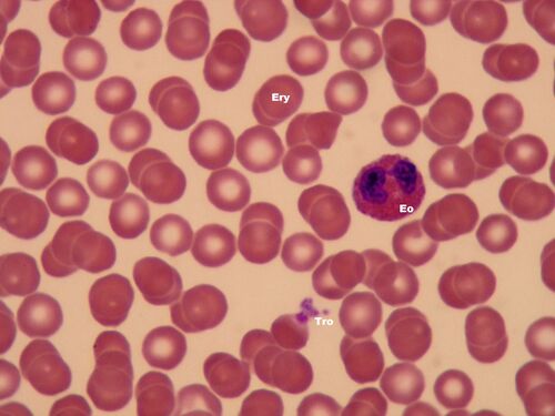
Name: Blood film slide - eosinophil (staining according to Pappenheim)
Popis: Eo – eosinophil with typical glasses-like nucleus, Ery – erytrocytes, Tro - trombocytes
Slide 3[edit | edit source]
Name: Blood film slide - basophil (staining according to Pappenheim)
Description: Ba – basophil (granules in cytoplasm completely cover the nucleus), Ery – erytrocytes
Slide 4[edit | edit source]
Name: Blood film slide - lymfocyte (staining according to Pappenheim)
Description: Ly – lymfocyte with nucleus and narrow cytoplasm in shape of thin sickle, Ery – erytrocytes,
Tro - trombocytes
Slide 5[edit | edit source]
Name: Blood film slide - monocyte (staining according to Pappenheim)
Description: Mo – monocyte with kidney-like nucleus, Ery – erytrocytes, Tro - trombocytes
Slide 6[edit | edit source]
Name: Blood film slide (staining according to Pappenheim)
Description: Neu – neutrophil (segmented nucleus), Eo – eosinophil (glasses-like nucleus and appearing red cytoplasm), Ery – erytrocytes, Tro - trombocytes
Slide 7[edit | edit source]
Name: Blood film slide (staining according to Pappenheim)
Description: Neu – neutrofil (segmented nucleus), Mo – monocyte (the biggest blood cell, kidney-like nucleus), Ery – erytrocytes, Tro - trombocytes
Slide 8[edit | edit source]
Name: Blood film slide – blood sample with immature neutrophils (staining according to Pappenheim)
Description: Ty - in Czech "tyčky" = immature neutrophils
Slide 9[edit | edit source]
Name: Capillary – electronogram
Description: 1 – erytrocyte in the transverse section (biscuit shaped), 2 – trombocyte, 3 – nucleus of the endothelial cell,
4 – cytoplasm of the endothelial cell, 5 - collagen fibrils in longitudinal and transverse section
Slide 10[edit | edit source]
Name: Eosinophil – electronogram
Description: 1 - nucleus, 2 – granules (dense center is so-called internum or marrow, light cover is so-called
externum or matrix)
Slide 11[edit | edit source]
Name: Lymfocyte – electronogram
Description: 1 - nucleus, 2 - thin rim of the cytoplasm
Slide 12[edit | edit source]
Name: Bone marrow – film slide (staining according to Pappenheim)
Description: K – blood cells – a mixture of different stages of development of the red blood cells, white blood cells and megakaryocytes,
T – fat cells


