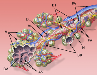
Bronchioles
On the surface of the lungs, small polygonal fields surrounded by tissue can be seen. These are lung lobules, i.e. sections of lung tissue ventilated by one bronchiole each. Bronchioles are behind the segmental bronchi in the branching hierarchy and further branch into terminal and respiratory bronchioles . During the branching of the bronchial tree , this "pipe" carrying air is simplified in its structure, and this is also reflected in the structure of the bronchioles:
- cartilage and seromucinous glands are completely absent in the wall from a diameter of approximately 1 mm
- the multi-rowed columnar epithelium with cilia and goblet cell (sometimes incorrectly referred to as the respiratory epithelium) gradually changes to the single-layered cuboidal epithelium. Cilia decrease, goblet and basal cells disappear and are replaced by Clara cells
Bronchioles[edit | edit source]
Unlike the bronchi, there are no lymph nodes, subepithelial glands, or continuous plates of cartilage of any shape.
Tunica mucosa[edit | edit source]
- Epithelial lamina
- the lumen of the widest part of the bronchiole is covered by the continuation of that in the bronchi, i.e.a multi-rowed columnar epithelium with cilia and goblet cells. It gradually decreases, goblet cells disappear and are replaced by Clara cells
- Lamina propria mucosae
- the fibrous layer consists of sparse collagenous tissue with smooth muscle cells
- due to the absence of cartilage, elastic fibers play a very important role here , which are connected to the elastic fibers around the alveoli, to the septa between the lobes and segments, and extend to the surface of the lungs. Together, they create a radially oriented tensile force that keeps the bronchioles open
Tunica muscularis[edit | edit source]
- thick layer of smooth muscle oriented spirally
Tunica adventitia[edit | edit source]
- a thin collagenous tissue in which blood and lymphatic vessels and nerves are embedded
Terminal bronchioles[edit | edit source]
Terminal bronchioles are the last division of the bronchial tree. The area of the lung parenchyma that is ventilated by 1 terminal bronchiolus is called the acinus pulmonalis .
Tunica mucosa[edit | edit source]
- Epithelial lamina
- the epithelium changes from single-layered cylindrical with cilia tosingle-layered cubic with cilia [1], in which Clara cells are scattered, the number of cilia also decreases
- basal cells have disappeared (it is already a single-layered epithelium), brush cells and DNES cells are rarely found here
- Lamina propria mucosae
- sparse collagenous tissue with smooth muscle cells and elastic fibers
Tunica muscularis[edit | edit source]
- thick layer of smooth muscle oriented
Tunicla adventitia[edit | edit source]
- a thin collagenous tissue in which blood and lymphatic vessels and nerves are embedded
Respiratory bronchioles[edit | edit source]
Respiratory bronchioles are the first structures where gas exchange and therefore respiration occurs . They are very similar in structure to terminal bronchioles, but alveoli protrude from their wall in places . Further, it branches several times;
Tunica mucosa[edit | edit source]
- Lamina epithelialis
- the lumen is lined by a single-layered cuboidal epithelium , initially ciliated, with scattered Clara cells. In the distal direction, however, the cilia disappear and Clara cells dominate more and more
- scattered brush cells and DNES cells can be found here and there in the epithelium
- Lamina propria mucosae
- a layer of sparse collagenous tissue rich in elastic fibers , isolated smooth muscle cells are arranged spirally
Tunica adventitia[edit | edit source]
- a thin collagenous tissue in which blood and lymphatic vessels and nerves are embedded
Links[edit | edit source]
Related articles[edit | edit source]
Respiratory system (histology)
Respiratory system lining cells:
Articles:
Bibliography[edit | edit source]
- KLIKA, Eduard, et al. Histologie pro stomatology. 1. edition. Praha : Avicenum, 1988. 448 pp.
- JUNQUEIRA, L. Carlos – CARNEIRO, José – KELLEY, Robert O. Základy histologie. 1. edition. Jinočany : H & H, 1997. 502 pp. ISBN 80-85787-37-7.
References[edit | edit source]
- ↑ PECKHAM,. Faculty of Biological Sciences, University of Leeds [online]. [cit. 2018-12-18]. <https://www.histology.leeds.ac.uk/respiratory/conducting.php>.


