Bronchus segmentalis SFLT
Bronchus - low magnification (hematoxylin-eosin staining)[edit | edit source]
Description: The mucous membrane of the bronchus is lined with a multi-row cylindrical epithelium with cilia. Underneath it is a loose collagenous tissue. In the wall we see hyaline cartilageand seromucinous glands.
Bronchus - multi-rowed cylindrical epithelium with cilia (hematoxylin-eosin staining)[edit | edit source]
Description:In stratified epithelium, all cells reach thebasal lamina,but their nuclei are located at different heights. It appears as if the cells form layers. Epithelium contains ciliated cells, basal cells [[goblet cell|goblet cells] and brush border cells. Mucin is stained blue in the goblet cell.
Intrapulmonary bronchus (staining - Alcian blue, hematoxylin-eosin)[edit | edit source]
Description:Hyaline cartilage in the bronchus is colored blue. A lymph node (follicle) can be seen in the wall above. The bronchus is surrounded by lung tissue (single-layered squamous epithelium).
Bronchus - multi-row cylindrical epithelium with cilia (hematoxylin-eosin staining)[edit | edit source]
Description: The stronger staining line represents the basal bodies of the cilia. Nuclei are stored in multiple rows, all cells are attached to the basal lamina.
Bronchus (hematoxylin-eosin stain)[edit | edit source]
Description: The wall of the bronchus consists of the mucosa (multi-rowed cylindrical epithelium with cilia and lamina propria - sparse collagen tissue), tunica fibro-musculo-cartilaginea, containing collagen tissue with an admixture of elastic fibers, smooth muscle and hyalinne cartilage. Externallytunica adventitia is found. Mixed sero-mucinous glands are found in the ligament in the wall of the bronchus. On the preparation, relatively wide outlets of these glands can be seen.
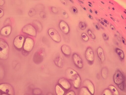
Bronchus - hyaline cartilage[edit | edit source]
Description: Chondrocytes are stored in lacunae, which are surrounded by intercellular matrix. On the surface of the cartilage we see perichondrium (collagenous tissue). The cells of the perichondrium differentiate into chondrocytes - they gradually become rounded.
Hyaline cartilage - bronchus (staining - alcian blue)[edit | edit source]
Description: Chondrocytes lie in lacunae. Several chondrocytes lying in close proximity form isogenetic group.


