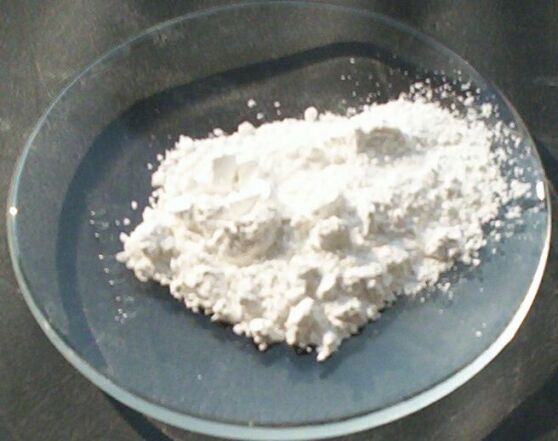
Calcium hydroxide
Calcium hydroxide is a white powder that is mixed with water or saline to form a paste. Meets most requirements for a root canal disinfectant insert.
Properties[edit | edit source]
- pH is 12-13 - a broad-spectrum antimicrobial agent
- high dissociation ability (75%) – the amount of hydroxyl ions around the insert is high
- low solubility (1.3 g/l) – long-term action is ensured
- biocompatible - no side effects
- support of remineralization
- high pH can be maintained for up to 30 days
- reacts with CO2 present in living tissues – formation of insoluble calcium carbonate and water, thereby creating a solid barrier preventing further penetration of hydroxyl ions into the surrounding tissue = self-limiting necrosis. During healing, the necrosis is pressed by a layer of newly formed ligament that calcifies and turns into dentinoid or osteoid tissue
- use in apexification, direct covering of the pulp, in induction of closure of the apical opening after vital extirpation
Indication[edit | edit source]
Disinfectant, antimicrobial canal liner[edit | edit source]
It is used in cases of:
- duct system is massively infected with bacteria - putrid smelling
- gangrene of the pulp
- channel open to DÚ for a long time
- extensive chronic periodontitis
- for time reasons
- due to complications
- disinfection of the more distant parts of the dentinal tubules and branches of the root canals, where the instruments and irrigation did not reach
We leave at least 7-10 days due to concerns about Str. faecalis (survives pH=11). If it drops to this value, the liner must be replaced after 10 days. Calcium hydroxide inactivates bacterial toxins through hydrolysis. Facilitates the effectiveness of sodium hypochlorite irrigation.
Therapeutic, remineralization supporting insert[edit | edit source]
It stops resorptive processes and potentiates mineralization around the tip of the root. It is possible to leave it for 2-3 months, the effectiveness is sufficient for the entire time. The application is carried out in the rinsed and dried canal using a spiral filler (lentulo). We introduce while running at slow speeds (max. 800/min) up to 2 mm from the apical constriction and then slowly extend for 30 seconds with constant rotation. For wide canals, injection is also possible. After application, we thicken it by sucking out the liquid with the opposite end of a paper pin and a ball of cotton wool, we remove the excess from the pulp cavity. After performing vital extirpation, even small remnants of the pulp in the apical part can hurt terribly after contact with hydroxide. Therefore, we apply the insert only to 1/3-1/2 of the length of the canal. Removal from the canal is easy - by rinsing with hypochlorite, or recapitulating, the most effective is the use of an endodontic ultrasonic tip with irrigation.
Indirect overlay[edit | edit source]
A field of softened dentin remains at the bottom of the cavity. If removed, there would be a risk of perforation of the pulpal wall and the need for endodontic treatment. Therefore, we will perform an indirect overlay. The execution depends on:
- pulp condition
- the size of a field of softened dentin
- the presence of bacteria in the dentin
Indication[edit | edit source]
- the pulp is healthy, possibly reversibly damaged, young (21–29 years)
- dentine area max 1 square mm
- dentin field (hard dentin, soft demineralized dentin, inner layer)
Procedure[edit | edit source]
Differentiation between demineralized and infected dentin is performed with the Caries detector. Application for 10s then rinse.
- demineralized dentin – unstained, capable of remineralization, collagen fibers intact
- infected dentin – stained is not capable of remineralization, the collagen fibers are already destroyed
We have to remove all the softened dentin except the pulpal wall. We prepare hard, absolutely healthy dentin on the gingival stage. We disinfect the cavity with:
- sparsely mixed Ca(OH)2 (we rinse a few minutes later)
- commercially produced Tublicid
- the most suitable 1-2.5% NaOCl on a cotton ball (4-5 minutes, rinse, dry)
Definitive Amalgam Filling
Carefully dry the cavity, apply solidifying Ca(OH)2 (about 2 mm). On top of it a resin-modified glass ionomer pad (Vitrebond, 3M ESPE, Fuji lining LC GC). Finally, a definitive filling made of amalgam.
Definitive Composite Resin Filling
Two types of treatment are used:
- omission of Ca(OH)2 - we directly apply the binding system (primer, bond), with the correct procedure, this cap is of better quality
- we etch enamel and dentin with acid, gently dry and then apply hardening Ca(OH)2, primer+bond and composite
Glass ionomer'
We apply Ca(OH)2 first, then GIC. 'We perform a vitality test every six months. Progressive caries removal
It is mainly performed for severely carious M1 in young patients.
- 1st visit: clinical examination, x-ray examination, vitality test, excavation of carious dentin. Leaving a small area of carious dentin near the pulp. I will apply non-hardening preparation Ca(OH)2 and make a temporary filling. Ca(OH)2 kills remaining microorganisms, neutralizes acids, hardens and dries softened dentin, induces formation of tertiary dentin.
- 2nd visit: vitality test, removal of temporary filling and pad, excavation of remaining dentin. Apply solidifying Ca(OH)2, pad, definitive filling.
Direct overlay[edit | edit source]
The task is also to preserve the vital pulp, we treat already opened pulp. Both dentin and odontoblasts are missing at the opening site (due to trauma) or by accidental opening of the pulp during dissection (in a hurry, with small-diameter tools in a high-speed elbow)
Terms[edit | edit source]
- healthy, vital pulp, young (up to 30 years)
- perforation diameter up to 1 square mm
- the edges of the perforation are in healthy dentin
It is necessary to ensure an aseptic operating field (cofferdam, we will replace drying, the patient must not rinse, suction)
We gently dry the dentin, we detect bleeding:
- at point opening – the pulp does not bleed or there is a small drop of light pink transudate, which is wiped with a cotton ball with 1-2.5% NaOCl, gently rinsed with a spray, dried.
- if light red blood - greater extent of perforation, then extirpation is necessary
- dark color of blood – probably irreversible inflammation, extirpation is necessary
Procedure[edit | edit source]
We apply solidifying Ca(OH)2 (about 2 mm) or without using Ca(OH)2 we apply the binding system and composite directly.
Forms of preparations of Ca(OH)2[edit | edit source]
- aqueous solutions (Hypocal, Calxyl)
- liner (Hydroxyline, Tubulitec)
- sealants (Gangraena Merz)
- cements (Dycal, Kerr-Life)
- light-curing preparations
- mixture with other materials (calcium salicylate cement + zinc oxide-eugenol cement)
Links[edit | edit source]
References[edit | edit source]
- PEŘINKA, Luděk. Základy klinické endodoncie. 2. edition. vydavatel, 2009. 0 pp. ISBN 978-80-903876-8-3.
- HELWIG, Elmar – KLIMEK, Joachim. Záchovná stomatologie a parodontologie. 1. edition. Grada Publishing, a.s, 1999. 0 pp. ISBN 80-247-0311-4.

