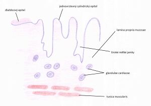
Cardia (histological slide)
From WikiLectures
The cardia of the stomach is a thin strip of muscle that represents the transition between the esophagus and the stomach and their mucosa. It consists of 4 basic layers, which are characteristic of the general structure of the alimentary canal.
Tunica mucosa[edit | edit source]
Epithelium[edit | edit source]
- transition of the stratified squamous epithelium of the esophagus into a single-layered cylindrical mucus-forming - gastroesophageal junction
Lamina propria mucosae[edit | edit source]
- sparse collagenous tissue, blood vessels, lymphatic infiltration
- glandulae cardiacae – branched tubular mucinous glands, the end parts of which can twist and have a spacious lumen
- - their secretory elements produce mucus and lysozyme
- - exceptions can be found that exclude HCl
- - structurally similar to esophageal cardiac glands
- the gastric glands open into wide and shallow pits (foveolae gastricae) that reach approximately 1/4 of the mucosa's height
Lamina muscularis mucosae[edit | edit source]
- bundles of smooth muscle cells
- usually an inner circular and an outer longitudinal layer
Tunica submucosa[edit | edit source]
- sparse collagenous tissue
Tunica muscularis[edit | edit source]
- 2 layers of muscle
- - inner layer - circular
- - outer layer - longitudinal
Tunica serosa[edit | edit source]
- sparse collagenous tissue
- on the surface of the mesothelium - a single-layered flat epithelium
Links[edit | edit source]
Related Articles[edit | edit source]
References[edit | edit source]
- MARTÍNEK, Jindřich and Zdeněk VACEK. Histological atlas. 1st ed. Prague: Grada, 2009, 134 pp. ISBN 978-80-247-2393-8.
- LÜLLMANN-RAUCH, Renate and Zdeněk VACEK. Histology. 1st Czech ed. Translation by Radomír Čihák. Prague: Grada, 2012, xx, 556 pp. ISBN 978-80-247-3729-4.
- Presentation from the website of the Institute of Histology, 1st Faculty of Medicine.
- JUNQUEIRA, L. – KELLEY, Robert – CARNEIRO, José. Basics of histology. - edition. H+H, 1997. ISBN 9788085787375.

