Cardiac wall arrangement, cardiac skeleton, chambers
Cardiac Wall Arrangement[edit | edit source]
The heart wall is composed of three distinct layers: the epicardium, myocardium, and endocardium.
- Epicardium: This is the outermost layer of the heart wall, also known as the visceral layer of the serous pericardium. It consists of mesothelial cells, adipose tissue, and connective tissue. The epicardium provides a protective layer and houses the blood vessels and nerves that supply the heart.
- Myocardium: The myocardium is the thickest layer and is composed of cardiac muscle cells called cardiomyocytes. These cells are responsible for the contractile function of the heart, enabling it to pump blood throughout the body. The myocardium's unique structure allows for strong, continuous, and rhythmic contractions.
- Endocardium: The innermost layer, the endocardium, lines the inner surfaces of the heart chambers and valves. It consists of endothelial cells and subendocardial connective tissue. The endocardium plays a crucial role in maintaining a smooth surface for blood flow and preventing blood clot formation.
Cardiac Skeleton[edit | edit source]
The cardiac skeleton, also known as the fibrous skeleton of the heart, is a dense, fibrous connective tissue structure located between the atria and ventricles. It consists of four fibrous rings (anuli fibrosi) and the membranous portions of the septa of the heart.
- Fibrous Rings: These rings encircle the orifices of the heart valves, providing structural support and attachment points for the valve leaflets. The rings include the left atrioventricular (mitral) ring, right atrioventricular (tricuspid) ring, pulmonary ring, and aortic ring.
- Trigones: The right and left fibrous trigones are interconnections between the fibrous rings and are the strongest parts of the cardiac skeleton.
- Functions: The cardiac skeleton serves several functions:
- Separates the atria from the ventricles.
- Maintains the valve orifices open.
- Provides attachment points for valve leaflets and cusps.
- Acts as an electrical insulator between the atria and ventricles.
- Provides a framework for the attachment of myocardial fibers.
Chambers of the Heart[edit | edit source]
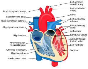
The heart is divided into four chambers: two atria and two ventricles.
- Atria:
- Right Atrium: Receives deoxygenated blood from the systemic veins (superior and inferior vena cavae) and pumps it into the right ventricle.
- Left Atrium: Receives oxygenated blood from the pulmonary veins and pumps it into the left ventricle.
- Ventricles:
- Right Ventricle: Receives deoxygenated blood from the right atrium and pumps it to the lungs via the pulmonary artery for oxygenation.
- Left Ventricle: Receives oxygenated blood from the left atrium and pumps it to the systemic circulation through the aorta.
The coordinated contraction and relaxation of these chambers ensure efficient blood flow through the heart and to the rest of the body.
References
Cardiac muscle tissue histology - Kenhub https://www.kenhub.com/en/library/anatomy/cardiac-tissue
Layers of the heart: Epicardium, myocardium, endocardium - Kenhub https://www.kenhub.com/en/library/anatomy/layers-of-the-heart
Fibrous skeleton of the heart: Anatomy and function - Kenhub https://www.kenhub.com/en/library/anatomy/cardiac-skeleton
The Heart - TeachMeAnatomy https://teachmeanatomy.info/thorax/organs/heart/
The Heart Wall - TeachMeAnatomy https://teachmeanatomy.info/thorax/organs/heart/heart-wall/


