Congenital defects of the gastrointestinal tract
Congenital atresias and stenoses of the digestive tract include:
- Esophageal atresia
- Congenital hypertrophic pyloric stenosis (pylorostenosis congenita)
- Atresia and stenosis of the small intestine
- Anal and rectal atresia
Other congenital causes of intestinal obstruction include:
- Superior mesenteric artery syndrome
- Malrotation of the intestine and volvulus
- Meconium ileus
- Megacolon congenitum
Esophageal atresia[edit | edit source]
Esophageal atresia is a developmental defect of the esophagus in which the esophagus is blindly closed and often fistula-connected to the trachea (up to 85%), creating a risk of aspiration. The incidence is 1:2000 to 1:4000, both sexes are affected equally. [1]:
Etiology[edit | edit source]
- failure of differentiation of the primary embryonic intestinal tube into the oesophagus, trachea, and lung
- may be part of the so-called VACTERL – developmental defects of the spine, anorectal region, heart, kidney, and radius (Vertebral anomalies, Anal atresia, Cardiovascular anomalies, Tracheoesophageal fistula, Esophageal atresia, Renal and/or Radial anomalies, Limb defects)
Classification[edit | edit source]
- according to Vogt (I–III) [2]:
- type I: a complete absence of the esophagus or a connective band instead of the esophagus, <1%
- type II: two distant stumps, no fistula absent, 8%
- type IIIa: upper esophagotracheal fistula, lower blind pouch, 1%
- type IIIb: lower esophagotracheal fistula, upper blind sac, most common (85-90%)
- type IIIc: upper and lower esophagotracheal fistula, 1%
- H-fistula: esophagus patent, H-shaped fistula present between esophagus and trachea, 5%
- according to R. E. Gross (A–E/H)
Clinical picture[edit | edit source]
- prenatally: polyhydramnios (and associated risk of preterm birth)
- postnatally: excessive salivation, cough at first drink, cyanosis, aspiration[1]
Diagnostics[edit | edit source]
- the gastric probe cannot be inserted
- chest and abdominal x-ray[1]
- X-ray contrast examination of the oesophagus – injection of aqueous contrast medium with a probe into the oesophageal stump, which in the most common type of Vogt IIIb atresia ends blindly at the level of Th2-4 vertebra
Treatment[edit | edit source]
- operative closure of the tracheoesophageal fistula – as soon as possible after delivery (risk of aspiration)
Congenital hypertrophic pyloric stenosis[edit | edit source]
Hypertrophic pyloric stenosis is acquired diffuse hypertrophy and hyperplasia of the smooth muscle of the pylorus and the entire stomach.
Etiology[edit | edit source]
The etiology is unknown; polygenic inheritance and environment are thought to be involved, and familial occurrence has been demonstrated in 15%. It is sometimes associated with hiatal hernia, esophageal atresia, or Turner syndrome. The incidence of the disease varies widely geographically, being approximately 1:5000 live births in the country, with up to five times more frequent occurrence in first-born boys. Newborns and infants between 3-6 weeks of life are affected, children older than 3 months rarely.
Clinical picture[edit | edit source]
- explosive vomiting in an arc dominates, projectile (up to 1 meter);
- vomit contains acidic gastric juices, the content is usually milk, the vomit is free of bile;
- dehydration occurs, the child has a large appetite, drinks eagerly; weight loss (dehydration, insufficient caloric intake)
- the child is lethargic, has constipation or hungry stools, has an aged appearance;
- can lead to severe hypochloremic alkalosis and hypokalaemia – severe condition, shallow breathing, loss of consciousness, convulsions – coma pyloricum;
- immediately after drinking, a peristaltic wave can be observed on the abdomen (from the left to the right epigastrium);
- on gentle palpation, in about 70% of children there is a palpable resistance = olive, the size of a cherry in the epigastrium, to the right of the midline – pyloric tumour;
- icterus may rarely occur
Laboratory examination[edit | edit source]
- typically hypochloremic alkalosis with hypokalaemia, hyponatraemia, and dehydration;
- hypochloremia can reach extreme values (below 75 mmol/l); its degree reflects potassium loss better than kalaemia;
- elevated gastrin values;
Diagnostics[edit | edit source]
- clinical picture
- abdominal ultrasound
- length (17 mm or more) and width (4 mm or more) of the pyloric canal are measured
- sensitivity 97%
- x-ray contrast imaging (GIT passage) is currently used when there is a diagnostic doubt
- gastric dilatation
- elongated and narrow pyloric canal (so-called rail or lace image)
- the contrast medium must be aspirated with a nasogastric probe after the examination (possible aspiration)
- rapid passage of contrast through the stomach eliminates pylorostenosis
- this examination may reveal other causes of vomiting without the presence of bile:
- gastric atony
- delayed gastric emptying,
- gastroesophageal reflux
Diferencial diagnostics[edit | edit source]
- other causes of explosive vomiting:
- intracranial hypertension,
- pyloric atresia,
- antral diaphragm,
- gastric duplication,
- gastric atony,
- delayed gastric emptying,
- gastroesophageal reflux;
- other causes of similar metabolic disruption:
- acute adrenal insufficiency – in hemorrhage or congenital hyperplasia – MAc,
- hyperkalaemia,
- loss of sodium in the urine;
- hereditary metabolic disorders – MAl can do disorders of amino acids metabolism (urea cycle disorder).
Treatment[edit | edit source]
- conservative management is not recommended
- treatment is surgical
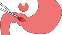
- most commonly, longitudinal pyloromyotomy of the hypertrophic pyloric musculature (Weber-Ramstedt operation) is performed:
- starting with a transverse laparotomy in the right epigastrium
- the straight abdominal muscles and the fascia of the oblique muscles are intersected longitudinally
- after opening the peritoneal cavity, the hypertrophic pylorus is luxated into the surgical wound
- a sharp longitudinal incision of the serosa and superficial muscle fibres is made (the incision starts 1-2 mm from the pyloroduodenal transition and ends at the pylorus to stomach transition)
- the muscle fibres are separated (along the entire length of the incision) by blunt dissection
- a complication is perforation of the mucosa – this should be sutured with absorbable material and covered with omentum
- patients with weight loss of more than 5%, metabolic alkalosis and hypochloremia should be parenterally rehydrated before surgery and the internal environment should be corrected within 24 hours
- most commonly, longitudinal pyloromyotomy of the hypertrophic pyloric musculature (Weber-Ramstedt operation) is performed:
- can also be performed by laparoscopic technique
- prognosis – good with early surgery.
Small intestine atresia and stenosis[edit | edit source]
Atresia and stenosis of the duodenum[edit | edit source]
The incidence of atresia and stenosis is 1:5000-10000. 30% of patients have Down syndrome and more than 50% of patients have associated birth defects.
Clinical course[edit | edit source]
Prenatally, polyhydramnios occurs due to interrupted amniotic fluid circulation. There is a so-called typical picture of two "bubbles", that are shut off by fluid. Postnatally with complete obstruction, a clinical picture of high ileus develops in the first 24 hours, which is manifested by violent vomiting with an admixture of bile. In 90%, the obstruction is located below the Vater’s ampulla. In the remaining 10% of cases, the vomitus does not contain bile. The physician may observe a bulging epigastrium, a sunken hypogastrium (boat-shaped abdomen), peristalsis is visible. Meconium does not go away. With partial obstruction, the clinical manifestation is late.
Diagnostics[edit | edit source]
Prenatally diagnosis is made by ultrasound screening. Postnatally, a native abdominal x-ray, which is typically depicted as two bubbles or levels, this applies to the stomach and dilated duodenum. In most cases, there is an absence of gas in the distal parts of GIT. More than 20 ml of fluid is aspirated from the stomach using a nasogastric probe. The normal volume of fluid in the stomach is 5 ml. This is followed by the insufflation of gas into the stomach, which allows the image of two bubbles to be reproduced on X-ray. A contrast X-ray of the upper GIT is performed to clarify the diagnosis and also to exclude malrotation or volvulus.
Treatment[edit | edit source]
The treatment is surgical, the patient undergoes a duodenoduodenal anastomosis.
Atresia and stenosis of the jejunum and ileum[edit | edit source]
The incidence of atresia and stenosis of the jejunum and ileum is 1:1500.
Clinical course[edit | edit source]
Prenatally polyhydramnios occurs. Postnatally the development of a clinical picture of moderate ileus occurs in the first 36 hours, resulting in vomiting with an admixture of bile. The abdomen is distended and respiratory distress – dyspnoea is present due to the high diaphragm. Meconium does not go away. Dehydration with hypochloremia and weight loss develop.
Diagnostics[edit | edit source]
Prenatally dilatation of the intestinal villi is evident on ultrasound. Postnatally a native abdominal X-ray is observed.
Treatment[edit | edit source]
Treatment is surgical, there is the removal of the atretic or stenotic section of the bowel and end-to-end anastomosis.
Anal and rectal atresia[edit | edit source]
Anal and rectal atresias are congenital closures or narrowings of the distal part of the intestine that result from failure of separation of the lower intestine from the ventrally located urogenital system during embryonic development. They are often associated with other congenital developmental defects (CDD), such as esophagus atresia, CDD of the urogenital system, lumbar and sacral spine. In addition, cardiac CDDs (ventricular septal defect) are common.
- Incidence 1:1500[1]
- 2 forms are distinguished:
- high atresia – the blind end is above the musculus levator ani (40% of cases);
- low atresia – the blind end is below the musculus levator ani (60% of cases).
Clinical course[edit | edit source]
- The anus is absent, mariscs are absent;
- this defect is usually detected in the delivery room when the newborn's body temperature is measured per rectum;
- if the defect is left untreated or unrecognised, ileus develops;
- due to fistulas with the urogenital tract, stool may pass, for example, through the vagina or urethra → severe infections of the uropoietic tract are the consequence.[1]
Diagnostics[edit | edit source]
- Postnatally: Ultrasound through the perineum;
- Fistulas can be visualized by injection of an aqueous contrast agent under skiascopy guidance.[1]
Treatment[edit | edit source]
- High atresia – colostomy, then corrective surgery at 3-5 months of age;
- Low atresia – transanal anoproctal plastic surgery (as soon as possible);
- incontinence is a long-term consequence.[1]
References[edit | edit source]
Related articles[edit | edit source]
Reference[edit | edit source]
Source[edit | edit source]
- BENEŠ, Jiří. Study materials [online]. ©2007. [cit. 2010-04]. <http://www.jirben.wz.cz/>.
Bibliography[edit | edit source]
- HRODEK, Otto – VAVŘINEC, Jan, et al. Pediatrics. 1. edition. Praha : Galén, 2002. 767 pp. ISBN 80-7262-178-5.
- ŠAŠINKA, Miroslav – ŠAGÁT, Tibor – KOVÁCS, László, et al. Pediatrics. 2. edition. Bratislava : Herba, 2007. 1450 pp. ISBN 978-80-89171-49-1.


