Contrasting in electron microscopy
Contrasting samples for TEM[edit | edit source]
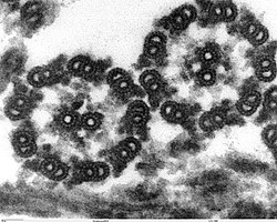
The resolution of samples in a transmissionelectron microscope depends on the scattering of electron beams upon impact on the sample. This dispersion depends on the atomic weight of the elements from which the specimen is made (the higher the atomic weight of the given elements, the higher the density will be reflected in the image). Biological structures themselves have a low contrast, because the biogenic elements (C, O, H, N) of which they are composed have small atomic weights, the values of which do not differ much from each other, so they are not discernible in an electron microscope.
There are many ways to increase the contrast of samples, for example, we can use the method of reducing the object aperture, reducing the accelerating voltage or increasing the thickness of the sample. However, these methods are not widely used because they lead to image blur. is mainly used to enhance contrast. Nowadays, absorption of heavy metal compounds onto biological structures.
The following metals are mainly used: U, W, Pt, Pb and Mn, also called electron dyes. These metals can increase the scattering of primary electrons and thereby increase the contrast of the sample structures.
"Positive" contrast method[edit | edit source]
Compounds of heavy metal bind to such surfaces that contain reduction sites (eg double bonds) and thus create high contrast structures that stand out against a less dense background.
- By incubation with vapors OsO4;
- double staining with uranyl acetate (uranyl acetate) and lead citrate, when uranyl acetate reacts mainly with nucleic acids and proteins and lead citrate then reacts with membranes proteins, nucleic acids and glycogen. After double-contrast, the sample thus obtains a uniform contrast of all cellular structures.
"Negative" contrast method[edit | edit source]
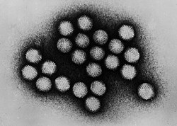
The method involves surrounding the sample with an electron-dense contrast agent containing heavy metal, which creates a dark background against which the lower-density sample will appear brighter. This method is suitable for observing small structures such as proteins a [[Polysaccharides, organelles, membrane systems, viruses and bacteria.
The most commonly used contrast agents are:
- AM = ammonium molybdate;
- UA = uranyl acetate;
- PTA = phosphotungstic acid solution.
Shading method[edit | edit source]
Shading consists in covering the preparation with a thin layer of metal, this ensures the highlighting of fine fibrillar structures and details of small macromolecules, e.g. collagenu, DNA, RNA, Ribosomes or cell wall. Metals with a high density (Au, C, U, Cr, W, Pt, Pd, Ni, Ge) are most often used. Plating is carried out in a high vacuum (10 -4 Pa) with the help of a vaporizing source of metal particles, when the heavy metal particles travel through the vacuum in a straight direction in different planes and accumulate on and around the sample. Where the sample is shadowed by the source, fewer metal particles will accumulate and this area will appear brighter - this is why rotary shading is already used today, where the sample rotates uniformly on a rotary platform during spraying. (In general, the rougher the surface of the preparation, the greater the angle of incidence of the sputtered metal we choose and vice versa.) The result of shading is a very fine granularity of metal particles, thanks to which fine structures are clearly visible.
Contrasting samples for SEM[edit | edit source]
The scanning electron microscope is mainly used for viewing the surface relief of samples up to 15×15 mm in size. It works on the principle of detecting reflected or ejected secondary electrons from the specimen. For observation, it is necessary that the preparations meet certain properties, such as being dust-free, stable in a vacuum, stable to the electron beam, sufficient thermal and electrical conductivity, preserved structure and volume, and minimal water content. Dried biological preparations do not meet these properties – they have low conductivity and when viewed in SEM without contrast, charging by the primary electron beam would occur, resulting in surface deformation and loss of image sharpness. The solution is to cover the preparation with a thin layer (10–20 nm) of a conductive material - most often a metal (Au, C, Pt, Pd, Ag), which will ensure protection of the preparation from thermal damage and at the same time increase the formation of secondary electrons, which will result in an increase in the signal (density) of the preparation.
This plating can be done in three ways:
By vacuum evaporation[edit | edit source]
When a high vacuum is created in the steaming apparatus and the metal is heated (electrically) to a temperature at which individual molecules begin to be released (evaporated) from its surface. These released metal molecules spread evenly in all directions through the vacuum and condense on the cooler specimen.
By ion evaporation[edit | edit source]
Which is based on the creation of rectified el. discharges (due to electric voltage) in a low-pressure argon atmosphere. The effect of electricity discharge will ionize the gas. The gas ions then travel to the cathode, around which there is a metal ring with which we want to dust the preparation. Accelerated gas particles eject metal particles from it, which spread evenly through the space of the vaporizer and collide with other gas ions. This creates a dense cloud of small metal particles in the entire space of the steamer, which uniformly covers the preparation with a thin layer.
Impregnation[edit | edit source]
Which is a chemical route that uses osmium or tannic acid. It is used in cases where vaporization equipment is not available, or when the preparation is partially conductive.
Links[edit | edit source]
Source[edit | edit source]
- NEŠČÁKOVÁ, M.: Principy kontrastování základních typů preparátů pro elektronovou mikroskopii, příklady, seminární práce pro předmět Histologické techniky bakalářského studia v oboru zdravotní laborant 2. lékařské fakulty Univerzity Karlovy. Praha: 2013
Literature[edit | edit source]
- NEBESÁŘOVÁ, J. Elektronová mikroskopie pro biology. kapitola 4.7: Kontrastování ultratenkých řezů, 2001. Dostupné z webové stránky: http://triton.paru.cas.cz/lem/
- NEBESÁŘOVÁ, J., Elektronová mikroskopie pro biology. kapitola 8.3: Pokovení preparátů, 2001. Dostupné z webové stránky: http://triton.paru.cas.cz/lem/
- ČECH, S., HORKÝ, D.. Přehled obecné histologie." 1. vydání. Skriptum. Brno: Vydavatelství Masarykova Univerzita, 2005. 140 stran
- Transmission Electron Microscopy (TEM) : sample preparation guide. Dostupné z webových stránek: http://temsamprep.in2p3.fr/accueil.php?lang=eng
- ŠAFÁŘOVÁ, K., Transmisní elektronová mikroskopie. Olomouc, 4. 12. 2009. Dostupnost z webových stránek: http://nanosystemy.upol.cz/upload/18/safarova_tem.pdf. prezentace pro Workshop: Mikroskopické techniky SEM a TEM. Centrum pro výzkum nanomateriálů, UP Olomouc
- Laboratoř elektronové mikroskopie. Metody přípravy biologických vzorků pro elektronovou mikroskopii aplikované LEM. Dostupné z webových stránek: http://web.natur.cuni.cz/~lem/index.php?p=metody
- ŠOBEROVÁ, T., Metoda negativního kontrastu pro diagnostiku virů a bakterií v transmisním elektronovém mikroskopu. České Budějovice: 2009. Dostupné z: https://theses.cz/id/miar9r/downloadPraceContent_adipIdno_12895. Bakalářská práce. Jihočeská univerzita v Českých Budějovicích. Zdravotně sociální fakulta
- Elektronová mikroskopie. Dostupné z webových stránek: http://old.vscht.cz/nmr/mol_model_bioinfo/lekce/mikroskopie.pdf
- Příprava preparátu pro elektronový mikroskop. Dostupné z: http://xarquon.jcu.cz/edu/zbb/elm.pdf
- Kontrastování („barvení“) ultratenkých řezů. Dostupné z: http://www.med.muni.cz/histology/MedAtlas_2/HP_txt8-2-4.htm
- Cytodiagnostické a imunocytochemické metody. Dostupné z: http://new.biologie.upol.czimunocytochemicke%20metody.htm
- Prozařovací elektronová mikroskopie (TEM). Dostupné z: http://new.biologie.upol.cztem.htm
- Rastrovací elektronová mikroskopie. Dostupné z: http://new.biologie.upol.cz
- KUBÍNEK, R., Moderní mikroskopie. Elektronová mikroskopie (TEM, SEM) Mikroskopie skenující sondou. Katedra experimentální fyziky Přírodovědecké fakulty, Univerzita Palackého v Olomouci
- MAŇÁKOVÁ, E., SEICHERTOVÁ, A., Metody v histologii, 1.vydání. Praha: Karilinum, 2002. ISBN 80-0230-X
- BEDNÁŘ, J., STANĚK, D., MALÍNSKÝ, J., KOBERNA, K., RAŠKA, I., Dnešní mikroskopie v biomedicíně. Pohyb molekul v přímím přenosu z živé buňky a další kouzla. Časopis VESMÍR č. 83, říjen 2004, dostupné z webových stránek: http://www.vesmir.cz
- JIRKOVSKÁ, M., Histologická technika pro studenty lékařství a zdravotnické techniky, 1. vydání. Praha: Galén, 2006. ISBN 80-7262-263-3
- HRDÝ, R., Elektronová mikroskopie (SEM, TEM, příprava vzorků, EDX, WDX). Budování výzkumných týmů a rozvoj univerzitního vzdělávání výzkumných odborníků pro mikro- a nanotechnologie (NANOTEAM) CZ.1.07/2.3/.00/09.0224. Instrukční a studijní materiál. Brno: 2011
- BOZZOLA, JOHN J., RUSSELL, LONNIE D., Electon Microscopy, Principles and Techniques for Biologists, Chapter 5 – Specimen Staining and Contrast Methods for Transmission Electron Microscopy. 2nd edition. Dostupné z webových stránek www.books.google.cz, Canada: Jones and Bartlett Publishers, 1999. ISBN-10: 0-7637-0192-0
- AYACHE, J., BEAUNIER, L., BOUMENDIL, J., LAUB, D., Sample Preparation Handbook for Transmission Electron Microscopy Techniques. New York: Springer. 2010. ISBN 978-1-4419-5974-4
- VAJNER, L,. Speciální metody v elektronové mikroskopii. Praha: Ústav histologie a embryologie, 2. lf. Materiál pro výuku
References[edit | edit source]
- Transmission Electron Microscopy (TEM) : sample preparation guide. Fig.3 [online]. [cit. 2013-11-07]. <>.
- ↑ HEJTMÁNEK,, M. a V. HORN. Vlas napadený kožní plísní - dermatofytem. [online]. [cit. 2013-11-07]. <>.


