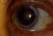
Corneal deposits
Arcus corneae senilis (gerontoxon)[edit | edit source]
Gray-white ring of corneal lipid deposits in limbus of the eye. Usually double-sided. It arises at the age of over 60. Its outer perimeter is sharply demarcated as opposed to the less sharply demarcated inner perimeter. Lipid deposits diffuse from the limbal vessels. When created before the age of 50, we look for lipid metabolism disorders (hypercholesterolemia) = arcus lipoides corneae. It does not affect visual acuity.
Kayser-Fleischer ring[edit | edit source]
Greenish discoloration of the cornea circumference caused by increased deposition of copper salts in the body. It is a symptom of Wilson's disease. Accumulated copper is deposited in the corneal limbus in the lamina limitans posterior (Descement's membrane), causing a circular olive green to brown color at the corneal-white border.
Argyrosis and chrysiasis[edit | edit source]
- Argyrosis - deposits in the deep layers of the cornea and on the conjunctiva for local medication containing silver salts.
- Chrysiasis - gold deposits of the periphery of the cornea for systemic application of gold (more than 1-2 g)
Cornea verticillata[edit | edit source]
Bilaterally centrally placed gray-brown deposits of corneal epithelium associated with long-term treatment with amiodarone or chloroquine. It does not affect visual acuity. It is also described as an ophthalmic manifestation of Fabry disease.


