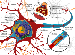
Dendrite
Dendrites (in Greek it means "tree") are branched protrusions from the neuron's soma that transmit post-synaptic potentials to it. They have high variability in the branching pattern and extent (characteristic for individual neuronal types): different numbers of axonal contacts (up to approximately 100 000) and different types of contacts (axo-shaft, axo-spine, dendro-dendritic). The dendrites contain dendritic organelles: neurofilaments, neurotubules, endoplasmic reticulum, mitochondria, ribosomes (metabolic autonomy). There are also special dendritic organelles: dendritic spines, dendritic swellings.
Dendrite's Special Functions[edit | edit source]
- Enlarged surface area to receive signals from axons of other nerve cells:
- The size of the dendritic tree limits how many synaptic inputs the neuron can receive.
- The orientation of the dendritic tree determines the types and number of sources from which it can receive synaptic connections.
- Transmission of received signals:
- Because dendrites are long, narrow, branching structures, the synaptic signal produced in the dendrites is significantly attenuated (due to increased resistance) by the time it reaches the soma. Thus they cannot propagate the action potentials.
- A dendrite may be considered to be an electrically leaky cable having a relatively low-resistance cytoplasm surrounded by a membrane consisting of resistive and capacitive elements in parallel. Therefore, the signal is conducted by electrotonic conduction: If a steady signal is applied to the end of a dendrite, the attenuation of the signal with distance will critically depend on the specific membrane resistance of the dendrite (the membrane potential will decline exponentially – decremental conduction).
- The farther the origin of excitation from the soma (cell body), the greater the degree of the decrement until the current reaches the cell body.
- Measurements made in vivo suggest, that neuron's cable properties are not fixed quantities. It appears more efficient than the mathematical models indicate. The most probable explanation is based on the presence of accumulations of the voltage-gated channels in various location of the dendritic membrane (at the heads and necks of the dendritic spines, at the dendritic branching points, and even at the whole dendritic segments).
- These accumulations (hot-spots) may recover the declining membrane potential and dramatically increase the effectiveness of conductance: pseudo-saltatory transmission by dendritic spines.
- Voltage-Gated-Channels at the dendritic membrane are selectively permeable to either Na+ or Ca2+. At the peripheral branches, the Ca2+ channels are more numerous, meanwhile the Na+ channels are present at more distal segments. As the Ca2+ channels operate in lower speed than the Na+ channels, the transmitting signal travels along the dendrite with a different speed.
- Elementary integrative function: summation of excitatory + inhibitory potentials before the final current reaches the cell body.
- Possible contribution to memory: facilitation is possible due to plasticity of dendritic spines.
- Possible source of neuromodulators and tissue factors.
Dendritic Spines[edit | edit source]
Dendritic spines are small outgrowth of the cell membrane of the dendrite. This is where a single synapse with an axon typically takes place.
Anatomy & Function of the Dendritic Spines[edit | edit source]
Dendritic spines are usually described by a bulbous head, connected via a thin cytoplasmic protrusion (neck) on the parent dendrite. Their length is maximum 2 μm and the spine head volumes can be 0.01 µm3 to 0.8 µm3. There can be thousands of dendritic spines on a neuron. The distribution density of dendritic spines ranges from 20 to 50 spines per 10 µm stretch of dendrite. A dendritic spine is a small membranous protrusion from a neuron's dendrite that typically receives input from a single synapse of an axon. Dendritic spines behave as a storage site for synapses and are responsible for collecting post-synaptic potentials and transmitting them to the parent dendrite. Most spines have a bulbous head (the spine head), and a thin neck that connects the head of the spine to the shaft of the dendrite.
The most notable classes of spine shape are:
- thin;
- stubby;
- mushroom;
- branched.
The variable spine shape and volume is thought to be correlated with the strength and maturity of each spine-synapse.
Electrical Properties[edit | edit source]
The conduction is done by electrotonic conduction (passive conduction of current). Due the spine's small size, the spine has a high input resistance. The spine's size is inversely proportional to the resistance. As like in any cable, the more space an electron has to travel through, the less resistance it will encounter in doing so. The synaptic potentials are relatively fast due to the relatively small capacitance of the spines, however the capacitance of the whole dendrite however becomes higher as the number of spines increases. Since the diameter (width) of the spine is much smaller than the diameter of the parent dendrite, this creates an impedance mismatch which eventually results in the spine following the potential of its parent dendrite.
Morphological Changes (Manifestations of Plasticity)[edit | edit source]
Dendritic spines have the advantage of plasticity. They can change their shape, volume and number within small time periods. These changes can result to change of capacitance and resistance. The actin cytoskeleton is main enabler of this plasticity, due to the inherent capability of actin remodelling. Spine maintenance and plasticity can be activity-dependent (ribosomal accumulation at the spine’s base) or activity-independent.
Links[edit | edit source]
Related articles[edit | edit source]
Sources[edit | edit source]
- Lecture Notes: Prof. MUDr. Jaroslav Pokorný DrSc.
Bibliography[edit | edit source]
- HALL, John E – GUYTON, Arthur Clifton. Guyton and Hall Textbook of Medical Physiology. 11. edition. Saunders/Elsevier, 2005. ISBN 0721602401.
- DESPOPOULOS, Agamnenon – SILBERNAGL, Stefan. Color Atlas of Physiology. 5. edition. Thieme, 2003. ISBN 3135450058.


