Development of oesophagus, stomach and small and large intestine
1. Formation of Gut tube:[edit | edit source]
Formation of gut tube and body cavity which occurs at the same time as neuralation. The epithelium and glands of the oesophagus are formed as of the 4th week from the entoderm of the foregut, connective tissue and muscle from the surrounding mesoderm. Since only the distal part of the foregut has its own artery in the Truncus coe- liacus, the oesophagus is supplied by various blood vessels in its various sections.
At end of third week Lateral plate mesoderm splits up into visceral (splanchnic) and parietal (somatic) layers with clefts appearing in the lateral plate.
a) Visceral layer – adjacent to the endoderm forming the gut tube folds ventrally becoming intimate with the gut tube and becomes continuous with visceral layer of extraembryonic mesoderm covering yolk sac. Visceral layer and its underlying endoderm make up the splanchnopleure.
b) Parietal layer – adjacent to surface ectoderm and continuous with extraembryonic parietal mesoderm layer of amnion. Parietal layer and its overlying ectoderm make up the somatopleure.
During 4th week sides of embryo grow ventrally forming 2 lateral body folds consisting of the somatopleure and cells from adjacent somite as these folds progress the endoderm layer also folds ventrally forming gut tube with the closure aided by growth of head and tail regions causing the embryo to curve into fetal position. Everything closes off of the ventral body wall except for region of connecting stalk which becomes the umbilical cord. The closure of gut tube is complete except for connection to midgut region called vitelline (yolk sac) duct.
The Primitive gut is now formed and is subdivided into three parts: 1) Foregut (from pharyngeal tube till liver bud) 2) Midgut (from liver bud to 2/3rd of right and 1/3rd of left colon) 3) Hindgut (from 1/3rd of left colon till cloacal membrane).
1.1 Foregut Development[edit | edit source]
The Epithelial lining of foregut and some of it's parenchyma is made of endoderm.
- Thyroid gland (follicular cells)
- Parathyroid glands
- Thymus
- Liver (hepatocytes)
- Gallbladder
- Pancreas (exocrine and endocrine components, including acinar and islet cells)
- Respiratory system (epithelium of the trachea, bronchi, and lungs)
- Esophagus (epithelial lining)
- Stomach (epithelial lining)
- Proximal duodenum (epithelial lining, up to the major duodenal papilla)[1]
1.1.1 Oesophagus[edit | edit source]
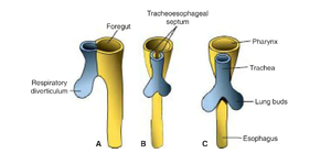
At around 4th week, respiratory diverticulum (lung bud) appears at ventral wall of the foregut at the border with pharyngeal gut. Tracheoesophageal septum parts this diverticulum from dorsal part of foregut.
As of the 4th–5th week, the oesophagus is separated by a tracheoesophageal septum from the lower respiratory tract, which also originates from the foregut
At first, the oesophagus is short but when heart and lungs descend it lengthens rapidly. Muscular coat is formed by visceral mesenchyme. It is striated in upper 2/3rd and innervated by vagus. The lower third is smooth and innervated by splanchnic plexus.
1.1.2 Stomach[edit | edit source]
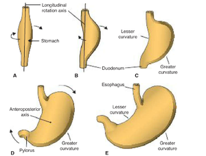
The stomach begins its development from the foregut in the fourth week as a fusiform dilation in close approximation to the respiratory diverticulum in the primitive thoracic region. The growth of esophageal region determines the position of the stomach. After lengthening of esophageal region the appearance and position of stomach changes greatly due to differing rates of growth in various regions of wall. Stomach rotates 90o around the longitudinal axis causing left side to face anteriorly and right side to face posteriorly hence left vagus nerve that initially innervated left of stomach now innervates anterior wall, same thing with posterior wall. At the same time, the original posterior wall of the stomach grows faster than anterior portion forming greater and lesser curvature. The Cephalic and caudal ends of stomach originally lie in midline but stomach roates around anteroposterior axis so that pyloric part (caudal) moves right and up and the cardiac (cephalic) portion moves to left and downward. The stomach is attached to dorsal body wall by dorsal mesogastrium and to ventral body wall by ventral mesogastrium which is part of septum transversum.
1.1.3 Small and Large Intestine[edit | edit source]
The small and large intestines develop from the midgut and hindgut, respectively. The small intestine arises from the cranial limb of the midgut's primary intestinal loop, which elongates and undergoes a 90° counterclockwise rotation during the 6th week as it herniates into the umbilical cord. By the 10th week, it retracts into the abdominal cavity, completing an additional 180° counterclockwise rotation for a total of 270°. The jejunum, ileum, and duodenum differentiate and become fixed to the posterior abdominal wall by mesentery. The large intestine originates from the caudal limb of the midgut (forming the cecum, appendix, ascending colon, and proximal transverse colon) and the hindgut (forming the distal transverse colon, descending colon, sigmoid colon, rectum, and upper anal canal). The cloaca divides into the anorectal canal and urogenital sinus during the 7th week, while the anal canal's distal segment develops from the ectoderm. The large intestine becomes fixed retroperitoneally, except for the transverse colon, which remains intraperitoneal.
References[edit | edit source]
Used Literature:
Langman's Medical Embryology, 14th Edition, by T.W. Sadler: Chapter 15
The Developing Human: Clinically Oriented Embryology, 10th Edition, by Moore et al.: Chapters on gastrointestinal development.
- ↑ "Langman's Medical Embryology," 14th Edition, by T.W. Sadler: Chapter 5 (Development of the Digestive System)


