Development of the amniotic and yolk sac, chorion
[1]Amniotic Cavity
- Amnion (amnos = lamb in Greek; agnus in Latin)
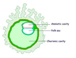
- Amniotic Cavity is a membranous sac encompassing and protecting the
conceptus in embryoblast.
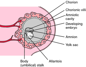
- The Amnion enlarges and encompasses the chorionic sac and covers the umbilical cord
- Day 8: cavities appear within the epiblast of
blastocystis bilaminaris (narrow slit between embryoblast a trophoblast) and the amnion is formed
-The Amniotic Sac is comprised of the Amniotic Cavity , Amniotic Fluid and is lined by amnioblasts(simple cuboidal epithelium) internally and externally by extraembryonic somatic mesoderm
Amniotic Fluid
-Initial Production: Amniotic fluid is initially produced by amnioblasts.
-Diffusion from Decidua Parietalis: It is then mainly produced by diffusion through the amniochorion from the decidua parietalis.
-Diffusion from Maternal Blood: Eventually, amniotic fluid is produced by diffusion from maternal blood in the intervillous spaces of the placenta.
-Fetal Respiratory and Digestive Tract: Amniotic fluid flows into the fetal respiratory and digestive tracts.
-Fetal Circulation: From 11 weeks, the fetus begins excreting urine into the amniotic fluid. By the end of the third trimester, the fetus swallows approximately 400 ml of amniotic fluid per day.
-Composition: Amniotic fluid is composed of 99% water.
-Function : Ensures symmetrical growth of conceptus
Acts as a barrier against infection
Enables movements of conceptus
Chorionic Cavity[1]
1. Trophoblast Layers during Implantation: The trophoblast differentiates into three layers during implantation: the syncytiotrophoblast, cytotrophoblast, and extraembryonal somatic mesoderm.
2.Formation of Extraembryonal Coelom: Cavities within the extraembryonal mesoderm fuse to form the extraembryonal coelom, also known as the chorionic cavity (cavitas chorionica).
3.Chorionic Cavity: The chorionic cavity is filled with fluid, providing a supportive environment for the developing embryo.
4.Connecting Stalk: The connecting stalk (pedunculus connectans) forms the connection between the embryo and the trophoblast, facilitating nutrient and waste exchange.
5.Role of Extraembryonal Mesoderm: The extraembryonal somatic mesoderm plays a critical role in the structural support and development of the chorionic cavity and connecting stalk.
Development of Chorion[1]
- Chorionic Sac and Cavity: The mesenchyma chorionicum forms the wall of the chorionic sac, while the mesothelium chorionicum lines the internal wall of the chorionic cavity.
- Lacunar Stage at End of 2nd Week: During the lacunar stage, the syncytiotrophoblast develops lacunae separated by trabeculae, which fuse into a labyrinth that becomes the future intervillous space.
- Formation of Primary Chorionic Villus: Cytotrophoblast columns invade the trabeculae, forming primary chorionic villi (villus primarius) and inducing adjacent extraembryonal somatic mesoderm.
- Development of Secondary Chorionic Villi: Starting from the 3rd week, primary chorionic villi develop further with the ingrowth of mesenchyme, forming secondary chorionic villi.
- Coverage of Chorionic Sac by 8th Week: By the 8th week, secondary villi cover the entire surface of the chorionic sac.
- Formation of Tertiary Chorionic Villi: Vessels develop within the mesenchyme of secondary villi, forming tertiary chorionic villi, which connect with the fetal circulation, facilitating nutrition and waste exchange.
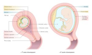
-Chorion Laeve Formation: The decidua capsularis compresses the villi in its vicinity, causing them to degenerate and form the chorion laeve (smooth chorion).
-Chorion Frondosum Formation: In the area of the decidua basalis, the villi multiply, resulting in the formation of the chorion frondosum (villous chorion).
- Differentiation of Chorion: The chorion laeve and chorion frondosum represent the differentiated regions of the chorion based on the interaction with different parts of the decidua.
Yolk Sac[1]
- The primary yolk sac develops from the extraembryonic coelem around Day 10/11, it is intially connected to the primitive gut however upon the growth of the chorionic cavity the yolk sac gets seperated and fades out
-The Secondary Yolk Sac develops on Day 13 from the extraembryonic mesoderm which also contributes to the formation of chorion and the connecting stalk
Functions
-Nutrient Transfer: In early development, the yolk sac is critical for the transfer of nutrients to the embryo before the placenta is fully functional.
-Hematopoiesis: It is the site of early blood cell formation (hematopoiesis) until this function is taken over by the liver and bone marrow.
-Germ Cell Origin: Primordial germ cells, which will eventually develop into the gametes (sperm and eggs), originate in the yolk sac and migrate to the developing gonads.
-Formation of Gut: It contributes to the formation of the primitive gut, as part of the yolk sac is incorporated into the embryo to form the gastrointestinal tract.
-Regulation of Development: The yolk sac synthesizes and transports proteins, and plays a role in the regulation of embryonic development through signaling pathways.


