Duodenum - divisions, positions, syntopy (draw scheme), blood supply
The small intestine is an organ located within the gastrointestinal tract. It is approximately 6.5m in the average person and assists in the digestion and absorption of ingested food.

It extends from the pylorus of the stomach to the ileocaecal junction, where it meets the large intestine at the ileocaecal valve. Anatomically, the small bowel can be divided into three parts: the duodenum, jejunum, and ileum.
Divisions and Syntopy[edit | edit source]
The most proximal portion of the small intestine is the duodenum. Its name is derived from the Latin ‘duodenum digitorum’, meaning twelve fingers length. It runs from the pylorus of the stomach to the duodenojejunal junction.
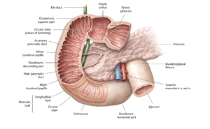
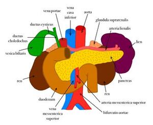
The duodenum can be divided into four parts: superior, descending, inferior and ascending. Together these parts form a ‘C’ shape, that is around 25cm long, and which wraps around the head of the pancreas.
- First Part (Superior Part):
- Begins at the pylorus and extends to the superior duodenal flexure.
- Positioned horizontally at the level of L1 (transpyloric plane).
- The initial 2 cm is intraperitoneal, forming the duodenal cap or ampulla, and is a common site for peptic ulcers.
- Relations:
- Anterior: Liver and gallbladder.
- Posterior: Portal vein, gastroduodenal artery, common bile duct.
- Superior: Hepatoduodenal ligament.
- Inferior: Pancreatic head.
- Second Part (Descending Part):
- Runs vertically along the right side of the vertebral column, extending from the superior to the inferior duodenal flexure.
- Crosses levels L1–L3.
- Contains the major duodenal papilla (opening of the hepatopancreatic ampulla of Vater) and the minor duodenal papilla (opening of the accessory pancreatic duct).
- Relations:
- Anterior: Transverse colon, right lobe of the liver, gallbladder.
- Posterior: Right kidney, ureter, renal vessels.
- Medial: Pancreatic head, bile duct.
- Third Part (Horizontal Part):
- Runs horizontally from right to left across L3, anterior to the aorta and inferior vena cava (IVC).
- Crossed anteriorly by the superior mesenteric vessels (artery and vein) and the root of the mesentery.
- Relations:
- Anterior: Superior mesenteric vessels, jejunum.
- Posterior: Aorta, IVC.
- Superior: Pancreatic head.
- Inferior: Jejunal loops.
- Fourth Part (Ascending Part):
- Ascends to the left of the aorta to the duodenojejunal flexure at the level of L2, where it transitions into the jejunum.
- Supported by the suspensory ligament of Treitz, which is important in distinguishing the upper from lower gastrointestinal bleeding.
- Relations:
- Anterior: Stomach.
- Posterior: Left psoas major, aorta.
- Superior: Pancreatic body.
- Inferior: Jejunum.
- First Part (Superior Part):
Positions[edit | edit source]
- Mostly retroperitoneal, fixed to the posterior abdominal wall.
- The first part (initial 2 cm) is intraperitoneal and mobile, connected to the liver by the hepatoduodenal ligament.
Blood Supply[edit | edit source]
- Arterial Supply:
- Supplied by branches of the celiac trunk and superior mesenteric artery (SMA), divided at the major duodenal papilla:
- Proximal (Foregut):
- Gastroduodenal artery → Superior pancreaticoduodenal artery.
- Distal (Midgut):
- Inferior pancreaticoduodenal artery (branch of SMA).
- Proximal (Foregut):
- Supplied by branches of the celiac trunk and superior mesenteric artery (SMA), divided at the major duodenal papilla:
- Venous Drainage:
- Veins mirror the arteries and drain into the portal vein, contributing to portal circulation.
- Lymphatic Drainage:
- Proximal part drains to the celiac lymph nodes.
- Distal part drains to the superior mesenteric lymph nodes.
References[edit | edit source]
- Standring, S. (ed.). Gray's Anatomy: The Anatomical Basis of Clinical Practice. 42nd ed., Elsevier, 2020.
- Schuenke, M., Schulte, E., Schumacher, U., et al. Atlas of Anatomy. 4th ed., Thieme, 2020.
Online Resources:
- Thieme Anatomy Atlases
- Radiology references on duodenal anatomy and imaging
- ↑ Schuenke, M., Schulte, E., Schumacher, U., et al. Atlas of Anatomy. 4th ed., Thieme, 2020.

