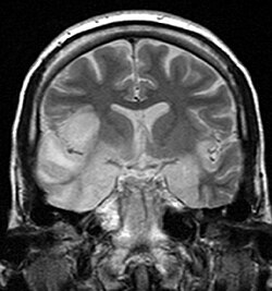
Encephalitis
Encephalitis means inflammation of the brain, caused mainly by a viral infection. However, it can also be caused by bacteria, parasites, etc. In many cases, not only the brain but also the meninges are damaged, so-called meningoencephalitis. Encephalitis is transmitted by arthropods, from other humans and from mammals.
Types of encephalits[edit | edit source]
Primary
- Caused by neurotropic viruses. These are transmitted to humans by animals. Neurons are damaged, plasma cells and lymphocytes infiltrate, and glia change. Typical intranuclear and cytoplasmic inclusions often occur in neurons .
Secondary
- In this case, brain injury occurs as a complication of the overall underlying disease. Viruses (eg enteroviruses, herpesviruses,…), rickettsiae, parasites ( malaria , toxoplasmosis ) , bacteria and spirochetes are also used here.
Viral encephalitis[edit | edit source]
In viral encephalitis, several typical features appear - edema, congestion, perivascular cuff infiltrates from macrophages, plasma cells, lymphocytes, varying degrees of neuronal damage, microglial nodules.
Rabies[edit | edit source]
Widespread disease worldwide. The causative agent is lyssavirus, contained in the saliva of infected animals. Viral changes in the animal's brain cause increased aggression and production of infected saliva, which is why it is most often transmitted to humans by biting. Clinically, only a local infection is present after the bite (often another pathogen of the animal's oral microflora). The time to clinical manifestation of rabies depends on the distance of the wound from the head (facial injuries for several days, limb injuries for several months). After entering the body, the virus travels along the axons of the peripheral nerves to the brain tissue.
Negri bodies are found in the cytoplasm of some neurons. Virions have been detected in the periphery of these bodies. The disease is treatable only in the latent phase before the development of clinical signs of CNS infection. After the onset of the disease, the lethality is almost 100%.
See the Rabies page for more information.
Poliomyelitis (Poliomyelitis acuta anterior, polio)[edit | edit source]
Foodborne infections caused by polioviruses. The virus first replicates in the mucosa of the oropharyngeal region and intestine, then enters the lymphatic vessels and blood. It shows an affinity for the motor area of the CNS gray matter. The biggest changes occur in the spinal cord (anterior horns). When motor neurons die, the relevant muscle groups atrophy, the limbs become immobile.
A complication of an acute infection is complete paralysis of the respiratory muscles. It also causes damage to the muscles, myocardium and PNS.
You can find more detailed information on the Polio page .
Herpetic Meningoencephalitis[edit | edit source]
The infectious agent is type 1 and 2 herpes simplex virus. It is the most common cause of viral meningitis in our country and in the USA. HSV-1 usually causes disease in adulthood (acute hemorrhagic-necrotizing encephalitis), HSV-2 disease is characteristic of the newborn (serous meningitis).
See Herpetic Meningoencephalitis for more information.
Tick-borne encephalitis (central european)[edit | edit source]
Transmission occurs by an infected tick (Ixodes ricinus). Exceptionally, transmission occurs through ingestion of infected meat or milk. The reservoir is small rodents. It occurs relatively often in the Czech Republic, usually with a benign course. We distinguish three forms: meningeal, meningoencephalitic, encephalomyelitic.
At first, vague flu symptoms appear. In a favorable situation, there is an overall improvement after a few days (the patient develops antibodies - there is no brain damage). In the worst case (about 3 to 7 days after the first symptoms), the problems get worse. Microscopically, we find perivascular mononuclear infiltrates in the venules and capillaries, as well as infiltrations in the soft diaper. Apoptosis and sometimes necrosis of neurons occur in both gray and white matter. The basal ganglia, cerebellum, elongated spinal cord and cervical spinal cord are most commonly affected .
See Tick-borne encephalitis for more information.
Uncommon viral encephalitis[edit | edit source]
- AIDS
- Encephalitis is present in about half of AIDS patients with HIV. It occurs in various forms ( aseptic meningitis , subacute encephalitis , vacuolar myelopathy (vacuolation of myelin sheaths).
- CMV
- Manifestations of encephalitis are also encountered in cytomegalovirus infection. It most often occurs in neonates of immunosuppressed patients. It affects any part of the spinal cord and brain, usually infecting the ependymus.
- Acute disseminated encephalomyelitis
- Also called acute disseminated leukoencephalitis. This infection can occur after smallpox vaccination and rabies vaccination. It is actually a T-cell mediated hypersensitivity reaction. It affects the white matter.
Slow viral encephalitis[edit | edit source]
As the name implies, these encephalitis are caused by slow viruses that have a very long latency period. This category includes:
- Subacute sclerosing panencephalitis
- It occurs in children and adolescents several years after measles. It leads to death within a few months to two years, with a progressively worsening picture of chronic meningoencephalitis. First there is a mental disorder, then loss of free movement, dementia, decerebratory rigidity. With a prolonged course, gradual atrophy develops, the consistency of the brain is stiff and the ventricles dilated.
- See subacute sclerosing panencephalitis for more information.
- Progressive multifocal encephalopathy
- Progressive rubella panencephalitis
Rickettsian encephalitis[edit | edit source]
The causative agent of this disease is Rickettsia prowazeki , which is transmitted by lice . In this case, encephalitis is part of the overall infection. Rickettsia attacks the endothelium and multiplies in the cytoplasm. The endothelium begins to necrotize, the lumen of the capillary is closed by a thrombus. Typical pericapillary nodules are formed in the capillary area. The oblongata, pons, basal ganglia and deep cortex are most often affected.
Fungal encephalitis[edit | edit source]
Encephalitis of this origin occurs exclusively in patients with immunodeficiency. The main mycosis is Candida albicans, Cryptococcus neoformans, Aspergillus fumigatus. Emerging thrombosis leads to heart attacks, granulomas and abscesses form.
Prion induced encephalopathies[edit | edit source]
They are characterized by the finding of spongiform vacuolization of neurons. The changes lead to the destruction of neurons and the proliferation of microglia and astrocytes. As far as the clinic is concerned, progressive dementia dominates, which is associated with pyramidal and extrapyramidal changes.
- Jakob-Creutzfeldt subacute spongiform ecephalopathy (SAD)
- A rare disease that occurs in adults. Organic psychosyndrome with paresis and dementia dominates. Pathological prions in neurons are produced. The cell cannot metabolize these prions, it accumulates in it and subsequently it is damaged. This infection is transmissible to other individuals.
- The Jacob-Creutzfeldt disease variant affects young people. Cortical plaques resembling Alzheimer's drusen form.
- Kuru
- It occurs in the natives of Papua New Guinea. It lasts for several years and ends with dementia. Earlier cannibalism played a major role in the transmission of the disease.
Encephalitis may also have an unclear etiology. We include Reye's syndrome. This is an acute encephalopathy that occurs in children (6 weeks to 16 years of age). The infection is associated with severe hepatic steatosis. Signs of brain damage may persist in the cured person. We can find edema in the brain without inflammatory changes.
Links[edit | edit source]
Related[edit | edit source]
- Meningitis • Meningitis (pediatrics)
- Viral meningitis • Serous meningitis and meningoencephalitis • Herpetic meningoencephalitis
- Purulent meningitis (infection) • Purulent meningitis (pediatrics) • Hemophilic meningitis • Tuberculous meningitis
- Infectious brain diseases • Neuroinfection, CNS / PGS inflammation
External links[edit | edit source]
References[edit | edit source]
- POVÝŠIL, Ctibor and Ivo ŠTEINER, et al. Special pathology. 2nd edition. Prague: Galén-Karolinum, 2007. ISBN 978-80-7262-494-2.
- MAČÁK, Jiří. General pathology. 1st edition. Olomouc: Univerzita Palackého, Lékařská fakulta, 2004. 345 pp. ISBN 80-244-0436-2.


