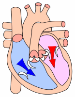
Events of the cardiac cycle
|
This article was marked by its author as Under construction, but the last edit is older than 30 days. If you want to edit this page, please try to contact its author first (you fill find him in the history). Watch the discussion as well. If the author will not continue in work, remove the template Last update: Monday, 08 Dec 2014 at 5.46 pm. |
The heart valves[edit | edit source]
Heart valves are fibrous tissues that separate the atria from the ventricles and the ventricles from large vessels that conduct blood to the pulmonary and systemic circulation. The function of the heart valves is to prevent blood backflow inducing a unidirectional flow of blood during the cardiac cycle events. This activity guarantees flow of a significantly large amount of blood to tissues and lungs enhancing the overall effect of systemic and pulmonary circulation.
- The atrioventricular valves - left mitral and right tricuspid - prevent regurgitation of blood into atria during ventricular systole. They are composed of membranous cusps that hang into the ventricles with their free endings being attached to the papillary muscles by thin tendinous threads called cordae tendinae, which prevent cusps from being pushed back into the atria during ventricular systole.
- The semilunar valves - aortic and pulmonary - are situated at bases of large arteries, preventing regurgitation of blood into the ventricles during the cardiac diastole.
Cardiac cycle[edit | edit source]
The cardiac cycle separates into two major phases, the ventricular systole and the ventricular diastole (which include the atrial systole). These events
Links[edit | edit source]
Bibliography[edit | edit source]
- HALL, John E. – GUYTON, Arthur C. Guyton and Hall Textbook of Medical Physiology. 12. edition. Saunders/Elsevier, 2010. ISBN 1416045740.
- Lecture Notes: Prof. MUDr. Jaroslav Pokorný DrSc.


