Examination of pituitary gland function
Physiology and pathophysiology of the pituitary gland[edit | edit source]
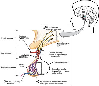
- The pituitary gland is an endocrine gland located on the cranial base and formed by two lobes with different ontogenesis, structure, function and regulation – anterior lobe (adenohypophysis) and posterior lobe (neurohypophysis).
- Pituitary gland is a stem associated with hypothalamus, which regulates pituitary function through hypothalamic hormones released into the capillary branches of the hypothalamic-pituitary portal system.
- Pituitary hormones have direct metabolic, cardiovascular and other effects on target tissues, but above all, through them the pituitary gland controls the function of peripheral endocrine glands – adrenal cortex, thyroid gland and gonads.
Adenohypophysis[edit | edit source]
- Anterior pituitary lobe (adenohypophysis) makes up 80% of the pituitary's weight.
- It is made up of glandular cells that synthesize and release into the circulation hormones of the protein structure – adrenocorticotropic hormone (ACTH), thyrotropic hormone (TSH), prolactine, somatotropic hormone (STH, growth hormone), endorphins, follicle-stimulating hormone (FSH) and luteotropic hormone (LH).
- The secretion of these hormones is regulated (stimulated or inhibited) by hypothalamic hormones (releasing and inhibiting factors).
- Adenohypophysis arises during ontogenesis from Rathke's protrusion of the posterior wall of the pharynx.
| Hormone | Secretory cells | Target tissue | Effects | Hyperfunctional and hypofunctional syndromes |
|---|---|---|---|---|
| Adrenocorticotropic hormone (ACTH) | Corticotropes | Adrenal cortex | Glucocorticoid secretion | Central Cushing's syndrome (Cushing's disease) |
| Beta-endorphine | Corticotropes | Opioid receptors | Suppression of pain perception | |
| Thyrotropic hormone (TSH) | Thyreotropyes | Thyroid gland | Thyroid hormone secretion | Central hyperthyreosis , hypothyreosis |
| Follicles stimulating hormone (FSH) | Gonadotropes | Gonades | Development and function of the reproductive system | |
| Luteotropic hormone (LH) | Gonadotropes | Gonades | Sex hormone secretion | |
| Somatotropic hormone (STH, growth hormon) | Eosinophil cells | Liver, adipose tissue | Growth, fat and lipid metabolism | Gigantism, acromegaly, dwarfism |
| Prolactine | Lactotropes | Ovaries, mammary glands | Lactation, affects estrogen and progesterone secretion | Galactorrea, gynecomastia |
Neurohypophysis[edit | edit source]
- The posterior lobe of the pituitary gland (neurohypophysis) is a functional and embryonic outbreak of the hypothalamus.
- It consists mainly of axonal endings of neurons in the supraoptic and paraventricular nuclei of the hypothalamus. These axons release the peptide hormones oxytocine and antidiuretic hormone.
- The posterior lobe also contains pituicytes, specialized glial cells related to astrocytes.
| Hormon | Secretory cells | Target tissue | Effects | Hyperfunctional and hypofunctional syndromes |
|---|---|---|---|---|
| Oxytocine | Nc. supraopticus and paraventricularis | Uterus, mammary glands | Uterine contractions, lactation | |
| Antidiuretic hormon (ADH, vasopresin) | Nc. supraopticus and paraventricularis | Kidneys, arterioles | Retention of water, vasoconstriction | SIADH, Diabetes insipidus |
Indications for examination[edit | edit source]
Endocrinological examination of the pituitary gland is indicated in case of clinical suspicion of:
- functional disorder,
- hypofunction = hypopituitarism, can be isolated, i.e., affecting one hormone, partial to complete, i.e., panhypopituitarism,
- hyperfunction (in most cases it is an overproduction of one hormone) – Cushing's syndrome, gigantism, acromegaly, prolactine,
- expansionary process in the area of the Turkish saddle (which is also often accompanied by a functional disorder),
- pituitary adenoma,
- craniopharyngeal, chordoma, meningioma, glioma or another type of tumor.
- Pituitary adenoma can be hormonally dysfunctional or hyperfunctional. The tumor itself can, by pressure on the surrounding tissue or by disrupting the hypothalamic-pituitary junction, induce either isolated or complete hypopituitarism, possibly diabetes insipidus.
- An example of a hormonally afunctional pituitary tumor is the so-called pseudoprolactine, which disrupts the transport of dopamine (prolactine-inhibiting hormone) into the pituitary by breaking the connection between the hypothalamus and the pituitary, with a consequent increase in prolactine secretion.
- Complications of pituitary tumors also include local invasion (damage to the visual tract, hypothalamic syndromes), hemorrhage, infarction, infection or malignant transformation.
- Autoimmune inflammation = pituitary – may also manifest as (pseudotumorous) pituitary expansion accompanied by a hormonal disorder (usually partial hypopituitarism). Swapping pituitary gland with adenoma can lead to incorrectly indicated surgery.
Examination procedure[edit | edit source]
The investigation procedure includes
- pituitary imaging methods (MRI or CT),
- hormonal examination – basal hormonal concentrations determined once or repeatedly during the day, functional (stimulation and inhibition) tests,
- biochemical and other laboratory tests in relation to presumed endocrinopathy (glycaemia, sodium, potassium, blood count, etc.),
- ophthalmological examination (especially visual field examination), possibly neurological examination and others.
Imaging Methods[edit | edit source]
Magnetic Resonance Imaging (MRI)
- Basic imaging method for suspected pituitary disease.
Comparison of MRI and CT in relation to pituitary examination
| MRI | CT | |
|---|---|---|
| Advantages |
|
|
| Disadvantages |
|
|
Hormonal examination[edit | edit source]
- exclude in the first place prolactinoma – unlike other hyperfunctional syndromes, there is primary medical treatment and not surgical treatment;
- rule out overproduction of other hormones – STH (acromegaly), ACTH (Cushing's disease), TSH (central hyperthyroidism); gonadotropins;
- diagnose hormonal deficits (hypopituitarism);
- exclude diabetes insipidus (anamnestic, possibly followed by a concentration test).
Prolactinoma[edit | edit source]
- Clinical picture:
- serum PRL concentrations – repeated intakes,
- response to dopaminergic agonists (therapeutic test) – in most cases the PRL level will decrease within a few weeks and later the tumor will regress,
- In differential diagnosis it is necessary to distinguish (true) prolactinoma from pseudoprolactinoma.
Acromegaly[edit | edit source]
- Clinical picture:
- serum STH concentrations basally – 3 samples at hourly intervals (due to circadian rhythm),
- serum IGF-I levels (single test),
- glucose inhibition test with determination of STH concentration.
Glucose Inhibition Test
- Test principle: hyperglycemia suppresses STH and ACTH secretion. After administration of 100 g of glucose orally on an empty stomach, the STH concentration drops below 1 μmol/l under physiological conditions.
Cushing's syndrome[edit | edit source]
- Clinical picture:
- free urinary cortisol – 24-hour collection,
- plasma cortisol – repeated intakes taking into account the circadian rhythm (or at least morning intake and intake at 11 pm),
- plasma ACTH,
- dexamethasone suppression test (usually a short test with a low dose of dexamethasone).
Dexamethasone suppression test[edit | edit source]
Basic test for suspected Cushing's syndrome.
Test principle: Inhibition test based on the principle of negative feedback. The application of a synthetic glucocorticoids (dexamethasone, DEX) leads physiologically to an attenuation of the ACTH-cortisol axis with a decrease in the secretion of both hormones. In patients with cushing's syndrome (either primary or secondary), this response is inadequate. The dexamethasone test has several variations of performance, the most commonly used of which is a short, „overnight“ with a single evening administration of a small dose of dexamethasone (1 mg or 2 mg per os). The ACTH and cortisol levels are compared at 2 blood collections – a basal collection in the morning before DEX administration and a collection in the morning after DEX administration.
An alternative is a six-day dexamethasone test, which combines a low (2 mg) followed by a high (8 mg) dose of DEX. A normal pituitary and adrenal response already occurs after a low dose of DEX. In patients with central Cushing's syndrome (pituitary adenoma), suppression occurs after a high dose of DEX. In the absence of a response, another type of Cushing's syndrome must be considered – peripheral or paraneoplastic Cushing's syndrome.
Stimulation test with corticotropin-releasing factor (CRF, 100 µg) with inferior sine petrosus catheterization
- The inferior petrosus sinus (SPI) is a venous raft that drains blood from the adenohypophysis. Local ACTH blood levels taken bilaterally from SPI (before and after CRF stimulation) are compared to peripheral blood ACTH levels.
- Use of the test in the differential diagnosis of the Cushing syndrome, differentiating between the pituitary microadenoma and paraneoplastic Cushing's syndrome, if the diagnosis was not possible by less demanding methods. In the case of paraneoplastic ACTH production (tumor production of ACTH outside the pituitary gland), there is no corresponding increase in ACTH levels in SPI after corticoliberin stimulation, because negative feedback predominates (peripheral tumor ACTH inhibits pituitary ACTH secretion).
Central hyperthyroidism[edit | edit source]
- Clinical picture:
- serum concentrations of TSH, free T4 and free T3.
Overproduction of gonadotropins[edit | edit source]
- Compared to previous hyperfunctional syndromes, it is much rarer.
- Clinical picture is not specific for diagnosis.
- LH, FSH, + men: testosterone.
Hypopituitarimus[edit | edit source]
- Demonstration of partial or complete disorders of adenohypophyseal hormones, possibly in combination with diabetes insipidus.
- hypocorticalism (plasma cortisol),
- central hypothyroidism (TSH, free T4),
- hypogonadism (LH, FSH, testosterone in men, menses in women),
- STH deficiency (STH in insulin stimulation test, IGF-I).
Insulin Hypoglycemic Test
- Test principle: Insulin-induced hypoglycemia stimulates the secretion of counterregulatory hormones, including ACTH, STH. Deficiency of ACTH and STH synthesis is manifested in the test by insufficient increase of their serum concentrations.
Methyrapone test
- Test principle: Metyrapone is a synthetic blocker of adrenal steroidogenesis. Blockade of cortisol synthesis by metyrapone stimulates ACTH secretion in the pituitary via feedback. ACTH synthesis deficiency is manifested by an insufficient increase within 120 minutes after metyrapone administration.
Stimulus tests using hypothalamic-releasing factors
- Principle of tests: Intravenous single application of releasing factors selectively stimulates the secretion of pituitary hormones.
It is often performed as a combined stimulation test with the simultaneous administration of several of the following factors, possibly in combination with an insulin test:
- thyrotropine-releasing hormone (TRH, 200 µg),
- gonadotropine-releasing hormone (GnRH, 100 µg),
- growth hormone-releasing hormone (GRF1-44, 100 µg).
Visual field examination (perimeter)[edit | edit source]
- Symptoms of oppression of the n. opticus by expansive process: scotomas, defects of the upper temporal quadrants, bitemporal hemianopia (bilateral failure of the lateral parts of the visual field), to blindness.
Links[edit | edit source]
Related articles[edit | edit source]
Literature[edit | edit source]
- MAREK, Josef. Farmakoterapie vnitřních nemocí. 2. edition. Praha : Grada, 1998. ISBN 80-7169-499-1.
- KRŠEK, Michal – HÁNA, Václav. Cushingův syndrom. 144. edition. Praha : Galén, 2006. ISBN 80-7262-399-0.
- KRONENBERG, Henry M.. Williams Textbook of Endocrinology. 11. edition. Philadelphia, USA : Saunders, 2008. ISBN 978-1-4160-2911-3.
- STÁRKA, Luboslav – ZAMRAZIL, Václav. Základy klinické endokrinologie. 2. edition. Praha : Maxdorf, c2005. ISBN 80-7345-066-6.
- ZAMRAZIL, Václav – PELIKÁNOVÁ, Terezie. Akutní stavy v endokrinologii a diabetologii. 1. edition. Praha : Galén, 2007. ISBN 978-80-7262-478-2.
- HÁNA, Václav. Endokrinologie : minimum pro praxi. 1. edition. Praha : Triton, 1998. ISBN 80-7254-000-9.
Portal:Pathophysiology
Portal:Internal medicine
Portal:Endocrinology


