Fluorescence in situ hybridization
|
This article was marked by its author as Under construction, but the last edit is older than 30 days. If you want to edit this page, please try to contact its author first (you fill find him in the history). Watch the discussion as well. If the author will not continue in work, remove the template Last update: Tuesday, 04 Dec 2012 at 10.08 pm. |
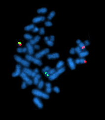
FISH (Fluorescence In Situ Hybridization) is a molecular genetic technique applied in cytogenetic examinations to detect small chromosomal rearrangements.
FISH - Technique: Basic steps[edit | edit source]
- Preparation of target DNA: DNA of metaphase or interphase cells are denatured into single-stranded DNA
- A DNA probe, corresponding to a specific chromosomal DNA sequence is labeled with a specific fluorophore
- Hybridization: Target DNA and the labeled DNA sequence are hybridized in situ to fixed metaphase or prometaphase chromosome spreads on a glass slide
- Each probe has the possibility of hybridizing specifically to two sister chromatids. The probe, marking a specific sequence of the chromosome is then visualized
Labeling[edit | edit source]
It is possible to either label the probe directly or indirectly. Direct labeling involves a labeled and modified nucleotide (often 2' deoxyuridine 5' triphosphate), which is directly incorporated into the probe. In indirect labeling, the DNA is labeled with a fluorophore to make the signal visible (not being incorporated into DNA). Indirect labeling can involve e.g. a hapten- or biotin- labeled probe which is then marked by fluorophore labeled antibodies or avidin.
Types of probes[edit | edit source]
- Centromeric (satellite probes)
- Locus specific probes
- Whole chromosome painting probes (e.g. used in mFISH)
Which chromosomal aberrations can be identified?[edit | edit source]
- translocations (balanced and unbalanced)
- copy numer changes
- additions
- deletions
- insertions
- inversions
- identifies chromosomal origins
- can identify specific p/q arms/bands
FISH vs. traditional Karyotyping[edit | edit source]
Traditional karyotyping allows scientists to view the full set of human chromosomes. Usually G-banding (Giemsa- stain) is used to display the bands of the chromosomes in a black and white pattern. Interpretation might be quite difficult, because the resolution is not always sufficient and usually an expert is needed, who might need a long time interpreting the bands.
FISH uses fluorescent dyes, which then can be painted with a specific computer program, so that even non-experts can easily see instances where e.g. a chromosome has parts of an other chromosome attached to it.
References[edit | edit source]
- STRACHAN, Tom, et al. Human Molecular Genetics. 4th edition. 2010. ISBN 978-0-8153-4149-9.
- PASSARGE, Eberhard. Color Atlas of Genetics. 3rd edition. 2007. ISBN 978-3-13-100363-8.


