General mechanisms of cellular damage
Each cell has a given range of structural and functional stability, which is conditioned by the genetically encoded program of differentiation , specialization and metabolism , the presence of neighboring cells and the availability of metabolic substrates . Under normal conditions, there is a dynamic balance in the sequence of metabolic events in which the cell is able to cope with the physiological demands, this state is referred to as normal homeostasis . A change in one of the parameters of this balance due to heightened stress or pathological stimuli leads to re-achievement of the balance by causing a change in another parameter. We refer to this process as adaptation, the result of which is a new equilibrium state.
Mechanisms of cell adaptation[edit | edit source]
B in the equilibrium state → B damaged → B adapted to the new equilibrium state
if the damage is too great:
B in equilibrium → B damaged → B permanently damaged → B disappearing
Causes of cell damage[edit | edit source]
1. Reduced oxygen supply[edit | edit source]
The result of reduced oxygen supply is hypoxia or ischemia .
Causes:
- impaired arterial inflow,
- clogging venous drainage,
- reduced capacity of blood to transport oxygen (anemia, CO poisoning, slowing of blood flow in cardiac failure). Oxidative phosphorylation in the mitochondria is mainly affected by hypoxia , and then anaerobic glycolysis is also blocked → radical reduction in production and lack of ATP.
2. Physical causes[edit | edit source]
Mechanical damage – trauma; temperature changes in both directions (frostbite, burns); sudden changes in atmospheric pressure; radiation – UV, ionizing and electrical damage
3. Chemical damage[edit | edit source]
A number of chemical agents work here . Medicines (ATB, cytostatics, etc.), physiological substances in toxic concentrations – O2, H2O, glucose, salts that disturb the steady state can also be toxic .
4. Genetic influences[edit | edit source]
They mainly cause growth disorders and a number of congenital malformations , including the effects of viruses on DNA - a direct cytopathogenic effect or the induction of an immune reaction against the given cell.
5. Immunological reactions[edit | edit source]
E.g. anaphylactic reaction to foreign proteins or antigens, or reaction to endogenous antigens, autoimmune diseases.
6. Nutritional imbalance[edit | edit source]
Lack or excessive supply of vitamins and trace elements .
General biochemical mechanisms of cell damage[edit | edit source]
There are several harmful agents whose action results in well-defined biochemical changes. The cell is particularly sensitive to glycolysis , the citrate cycle and oxidative phosphorylation .
Lack of oxygen and toxic effects[edit | edit source]
Critical oxygen deficiency manifests itself in two ways.
- A reduced oxygen level primarily means a loss of oxidative phosphorylation of ADP, and thus a reduced generation and subsequent depletion of ATP .
- Reperfusion damage by the formation of ROS – reactive oxygen species, which in biological systems serve as signaling molecules for gene expression, but due to their instability, damage lipids, proteins, and nucleic acids. Damage to macromolecules occurs because the mitochondria fail to switch back to oxidative metabolism and begin to form radicals .
ATP depletion[edit | edit source]
Adenosine triphosphate as a substrate for all types of ATPases for the reaction ATP = ADP + P+e .
It is generated by:
- by oxidative phosphorylation of ADP – reduction of oxygen electron transfer systems in mitochondria;
- glycolysis – ATP molecules are obtained in the absence of oxygen by utilization of glycogen, glucose and derivatives.
- Depletion below 5-10% will cause damage to critical cellular systems.
- It will reduce the activity of Na + /K + -ATPase → diffusion of potassium from the cell and rise of IC sodium level → IC edema – moving away of ribosomes from ER – blocking of proteosynthesis.
- Impairment of Ca 2+ -ATPase – maintains differential calcium concentration between cell and EC fluid.
Intracellular calcium level, calcium homeostasis[edit | edit source]
The Ca 2+ balance between the cell and the extracellular fluid as well as between intracellular compartments is maintained by the calcium ATPase (Ca 2+ -Mg 2+ -ATPase). This pump must be adapted to handle the huge concentration differences. The intracellular concentration of Ca 2+ in the cytosol is 0.1 μmol, while extracellularly it approaches up to 1.3 millimoles. Increased concentration of IC calcium is highly toxic! An increase in the IC level leads to ischemia or toxic damage , whereby calcium enters the IC environment from the EC environment or from compartments (e.g. leakage from the mitochondria and from the ER into the cytoplasm).
- Pathogenesis : increases membrane permeability and activation of some enzymes – ATPase – acceleration of depletion, phospholipase – damage to cell membranes, endonuclease , DNA-zyme .
Mitochondrial dysfunction[edit | edit source]
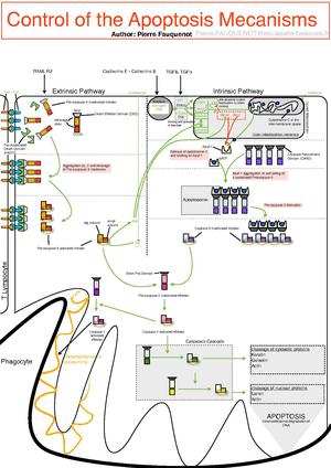
Mitochondria are very sensitive to a number of pathological attacks such as hypoxia , chemical damage , radicals , toxins , increased Ca 2+ in the cytosol → release of cytochrome c induction of apoptosis
Chemical damage to the cell[edit | edit source]
Cell poisons : As, CN, Hg, insecticides, herbicides, asbestos, CO, drugs, alcohol
Mechanisms of action :
1. direct connection to the molecular components of cell organelles
- E.g. HgCl 2 binding to -SH groups of proteins, CN – inhibition of cytochrome oxidase by binding to heme iron.
2. other chemicals, especially liposoluble, are biologically inactive, but converted into reactive toxic metabolites – most often ROS, RNS .
- It causes lipoperoxidation, the formation of cross-links between proteins (via -SH groups), DNA damage - by reacting with thymine DNA, they create single-stranded sections of DNA.
- Removal by antioxidants and enzymes: SOD (superoxide dismutase), glutathione peroxidase, catalase.
- These antioxidants decrease with aging → worse defense against oxidative stress and most likely the reason for death!
Disorders of membrane permeability[edit | edit source]
All of the above causes of cellular damage result in damage to membrane systems. The plasma membrane can also be damaged by bacterial toxins , viral proteins , complement components and lymphocyte products ( perforins ). Loss of barrier function (fence function) as well as loss of permeability of membrane cell systems (gateway function) is a prerequisite for the onset of irreversible cell damage .
Reversible and irreversible cell damage[edit | edit source]
Metabolic pathways that lead to cell damage combine with each other. The starting point of the following model of reversible and irreversible cell damage is lack of oxygen.
Reversible cell damage[edit | edit source]
- Reduced oxygen tension → loss of oxidative phosphorylation of ADP → reduced generation of ATP. After the breakdown of the oxidative phosphorylation system, the cell can generate ATP by switching to glycolysis – but with reduced yield. The subsequent accumulation of metabolites of glycolysis leads to an increase in osmotic pressure in the cell and increased lactemia with a decrease in pH. After glycogen stores are depleted , the level of glycolysis decreases rapidly.
- Collapse of the ATP-dependent sodium pump and increase in osmotic pressure → diffusion of potassium from the cell and accumulation of sodium and water in the cell → cell swelling, ER dilation, ribosomes moving away from the ER membranes → decrease in proteosynthesis, violation of the calcium pump → Ca 2+ into the cytosol .
Irreversible cell damage[edit | edit source]
- Here there is severe damage to the membranes and changes in their permeability → swelling and gradual decomposition of mitochondria and lysosomes. Increased flow of calcium into the cell → activation of proteases and endogenous phospholipases and thus loss of membrane phospholipids. Fission products – ''free fatty acids'' and acyl-carnitine change the permeability of mitochondrial and lysosomal membranes, which results in:
- leakage of mitochondrial dehydrogenases and cytochrome from mitochondria,
- swelling of lysosomes and rupture of their membranes with leakage of lysosomal enzymes into the cytoplasm and destruction of other cellular structures.
- These changes lead to the loss of proteins, enzymes, coenzymes and RNA through the hyperpermeable membrane and further to the increased expression of adhesion molecules by hypoxic cells. Adhesion molecules immigrate to leukocytes and the resulting inflammation will contribute to further cell damage. The result is final extinction .
links[edit | edit source]
Related articles[edit | edit source]
References[edit | edit source]
- POVÝŠIL, Ctibor. Obecná patologie. - vydání. Galén, 2011. 290 s. ISBN 9788072627738


