Hemoglobinopathy
Hemoglobinopathy is one of the most common hereditary diseases in the world. They are characterized by either a change in the formation of the entire globin chain or synthesis of an abnormal globin chain. An increased incidence of thalassemia and sickle cell disease is observed in areas with malaria. Heterozygous forms provide some protection against malarial infection.
Mutations of Hemoglobin[edit | edit source]
Mutations of the beta gene affect almost 50% of hemoglobin chains in heterozygotes; Alpha gene mutations affect only 25% of molecules, but they manifest before birth. Hereditary diseases caused by a disorder of hemoglobin production (hemoglobinopathy) can be divided into:
- diseases with a disorder of the globin chain structure;
- causing hemolytic anemie – sickle cell disease, unstable hemoglobin;
- reducing the ability of hemoglobin to carry oxygen - methemoglobinemia;
- diseases with a disorder of globin chain synthesis - thalassemia.
Hemoglobinopathies were the first inherited diseases to be diagnosed with DNA.
Mechanism of hemoglobinopathy:
- About 250 different structural mutations of hemoglobin are described - ⅔ of them affect the β-chain, less than ⅓ α-chain.
- Single base substitution - change in AMK in hemoglobin (HbS) structure.
- Two substitutions in different codons - change two AMK (HbC Harlem)
- Inequal crossing-over - eg Hb-Lepore - non-α-chain was created by recombination of the first part of the DNA delta-chain and the second part of the β-chain.
Diseases with a change of structure of hemoglobin chain[edit | edit source]
Disorders of hemoglobin structure causing hemolytic anemia[edit | edit source]
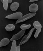
Sickle cell anemia - this is a severe autosomal recessively inherited hemolytic anemia, associated with thrush disorders, spleen dysfunction and so-called vasoocclusive crises - these are caused by capillary obstruction of the erythrocytes in the limbs, spleen and lungs. Without proper medical care, the disease is lethal. Heterozygotes are clinically healthy (or have very few clinical manifestations), and only part of the erythrocytes show sickling on examination.
The mutation is caused by a change in AMK in a polypeptide in the β-globin chain (point mutation). Valine ranks sixth instead of glutamic acid. We refer to altered hemoglobin as HbS. The cause of the confusion is a change in one nucleotide (A – T) in the GAG triplet, which causes a change in the isoelectric point of hemoglobin (instead of the negatively charged glutamic acid, there is a nonpolar valine). As the oxygen partial pressure decreases (i.e., deoxyhemoglobin is formed), HbS molecules aggregate into rod-shaped polymers, reducing plasticity and increasing erythrocyte fragility. Sickle-shaped erythrocytes pass worse through capillaries, can clog them and cause local hypoxia and heart attack. As it passes through the lungs and oxygenates, the shape adjusts again, but over time, the pathological sickle-shaped shape (irreversibly sickle-shaped erythrocytes) fixes.
Consequences:
- blood stagnation – hypoxia;
- aggregation of blood cells in circulation - ischaemia and infarcts;
- increased destruction of blood cells in the spleen – anemia.
Clinical Manifestations:
- hemolytic anemia with increased reticulocyte count, jaundice and a tendency to cholelithiasis;
- various symptoms (acute and chronic) resulting from ischemia (neurological symptoms - motor and sensory disorders; vision disorders, pulmonary microinfarcts - chest pain, pulmonary hypertension, muscle pain, aseptic necrosis of the femoral head, papillary necrosis of the kidneys with hematuria, skin ulceration… );
- Crises are sudden bouts of severe, long-term bone and muscle pain that are often caused by high-altitude stay, impaired lung function, increased metabolic activity, and dehydration.
Treatment:
Early diagnosis is needed to effectively treat and minimize complications. Newborn screening is therefore in place in some of the Middle East and US countries.
- Antibiotic prophylaxis (greater vulnerability to infections due to spleen dysfunction).
- Blood Transfusion
- Hematopoietic stem cell transplantation (bone marrow).
- Pharmacologically hydroxycarbamide (hydroxyurea) - increases gene expression of HbF (fetal hemoglobin - α2γ2), which does not contain defective β-subunits.
- Gene therapy - was described (2017) using a lentiviral vector (however, its widespread implementation in areas of the main occurrence in developing countries is unrealistic for financial reasons).
This disease is rare in our country, but the high frequency of the HbS allele is in many parts of the world - the Mediterranean, Arabia, India and especially sub-Saharan Africa (Nigeria, Democratic Republic of Congo and Tanzania); affects the black population in the USA (up to 8% frequency) In malarial areas, it is possible to observe positive selection for heterozygotes - they are more resistant to this protozoal infection. This is probably due to the reduced ability of Plasmodia to multiply within sickle cell erythrocytes and also to the increase in the total number of red blood cells. From a genetic point of view, the coherence with polymorphisms in genes related to HbF gene expression (HGB2, BCL11A, ...) is interesting, as a result of which the level of HbF is higher (up to 10%).
Hemoglobin C (HbC) - lysine is included in the 6th position in the β-chain instead of glutamic acid. The mutation is due to the exchange of guanine for adenine (GAG - AAG). HbC is less soluble and crystallizes in erythrocytes; the symptoms of hemolytic anemia are identical to sickle cell disease.
Hemoglobinopathy Affecting the Ability of Hemoglobin to Carry Oxygen[edit | edit source]
These mutations are an example of the mechanism of change in protein function. Stability and hemoglobin synthesis are not affected by these mutations
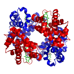
Methemoglobines (HbM) – the mutation affects the binding site of globin and heme, the so-called heme pocket. This affects the action of reductases on the binding of iron and oxygen (mutation leads to a change in the oxidation number of Fe2 + to Fe3 +, which does not bind oxygen). Physiologically, HbM is produced to a small extent but is reduced by methaemoglobin reductase. In HbM Hyde Park, AMK Histidine Tyrosine is replaced at position 92 in the β-globin chain. The result is a stronger binding of oxygen to iron and a slow release of oxygen by reductase in the tissues. Heterozygotes are cyanotic but without signs of oxygen deficiency. Homozygotes are not known, the combination is probably early lethal.
Short-term methaemoglobinaemia can occur in people with HbA after nitrate poisoning; Infants in whom reductase is not yet active are particularly at risk.
Diseases with impaired globin chain synthesis - thalassemia[edit | edit source]
Thalassemia is the most common monogenic hereditary disease (AR). It is a heterogeneous group of diseases with a disorder of α-globin chain synthesis (α-thalassemia) or β-globin chain (β-thalassemia). The second strand is synthesized in normal amounts and because it is in relative excess, it precipitates in erythrocytes and causes their premature destruction and exacerbates hypochromic anemia. Thalassemia heterozygotes are more resistant to malaria
α-thalassemia[edit | edit source]
The disease is caused by a disorder of α-globin chain production, the production of fetal and adult hemoglobin is impaired. Instead of HbA, hemoglobin is formed from four gamma chains or four beta chains - these tetramers are unable to carry oxygen. Anemia is microcytic, hypochromic, with extravascular hemolysis and splenomegaly, in severe cases, the fetus suffers from a lack of oxygen, it develops hydrops and other damage.
The most common cause is deletion; the presence of two identical α genes on chromosome 16 increases the likelihood of an abnormal synapse; the subsequent crossing-over results in a triplication of the α gene on one chromosome and a deletion on the other. There are two genes (four alleles) in the genotype for the α-globin chain, the degree of severity of the disease corresponds to the number of affected alleles:
- deletion of one allele is without clinical signs;
- deletion of two conditions minor thalassemia (possibly trans form A− / A−, or cis form −− / AA) and mild anemia,
- deletion of 3 alleles - severe hemolytic anemia with the formation of unstable hemoglobin HbH (β4), which precipitates on the membrane and cytoskeleton of erythrocytes as Heinz bodies;
- deletion of four lethal alleles already during intrauterine development, Bart's hemoglobin (γ4) or HbH (β4) may be formed.
β-thalasemia[edit | edit source]
β-thalassemia also causes anemia, but only after 3 months of age, at a time when HbF (γ-chain) synthesis is replaced by HbA (β-chain) synthesis. Excess α-chains damage erythroid cells and they break down already in the bone marrow (inefficient erythropoiesis), with a complete lack of β-chains, HbF (α2γ2) and HbA2 (α2δ2) occur in the blood. There is an increased amount of HbF and HbA2 in erythrocytes, as the production of γ and δ chains is not affected.
The β-chain may be insufficiently synthesized (β + thalassemia) or not produced at all (β0 thalassemia). The β-chain is encoded by 5 genes on chromosome 11.
β-thalassemia can be caused by all known disorders of the proteosynthesis mechanism: promoter mutation (mild BT, Japan), deletion of part of the β-globin gene (severe BT, Indians), chain fusion (severe BT Lepore, Italy), mutation of the mRNA splice site and new splice site (heavy BT, Africa), cap site mutation (light BT, Asia), mRNA end polyadenylation disorder (mild BT, Africa), stop codon generation (heavy BT, Mediterranean) or reading frame shift (heavy BT, Indians).
Anemia is microcytic, hypochromic, marked hepato- and splenomegaly, bone marrow hyperplasia. Repeated transfusions of erymass are necessary for the treatment (not only to replenish hemoglobin, but also to reduce erythropoiesis stimulation), but there is an excess of iron - the formation of oxygen radicals damaging various organs (liver - cirrhosis, pancreas - DM, myocardium - fibrotic changes).
References[edit | edit source]
Related Articles[edit | edit source]
- Structure of hemoglobin
- Hemoglobin and its derivatives
- Anemia
- Hemolytic and corpuscular anemia
- Hemolytic extracorpuscular anemie
External Links[edit | edit source]
Source[edit | edit source]
- ŠTEFÁNEK, Jiří. Medicína, nemoci, studium na 1. LF UK [online]. [cit. 11. 2. 2010]. <http://www.stefajir.cz>.
- PASTOR, Jan. Langenbeck's medical web page [online]. [cit. 12.4.2010]. <http://langenbeck.webs.com>.
- NEČAS, Emanuel. Patologická fyziologie orgánových systémů : part I. 2. edition. V Praze : Karolinum, 2009. 379 pp. pp. 63. ISBN 978-80-246-1711-4.


