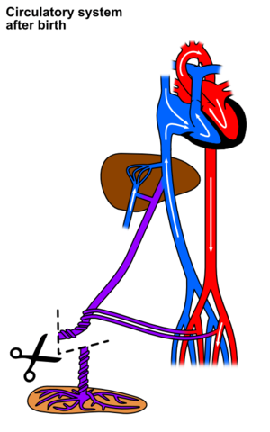
Human Fetal Circulation
Fetal circulation is the circulation of blood through the cardiovascular system in the human fetus. It's well known that the blood circulation in the humans varies. When the embryo develops into the fetus, it creates a functional cardiovascular system that cooperates with the mother's system. During birth, there are functional physiological changes that transform the shared system into an individual one for the fetus.
Fetal circulation is markedly different from circulation in adults. In the fetus, the main filtration site for plasma nutrients and wastes is the placenta, which is outside of the body cavity. In adults, the circulation occurs entirely inside the body.
Circulation in the Fetus Cardiovascular System[edit | edit source]
From the placenta the blood is 80% saturated with oxygen and enters embryo through the umbilical vein, located in the umbilical cord.
The blood flows in the direction to the inferior vena cava, passing the liver, where at its level enters ductus venosus.
In the liver, a small amount of blood supplies it by entering liver sinusoids, while the bigger amount continuous to inferior vena cava.
In the inferior vena cava the placental blood mixes with deoxygenated blood from lower limbs and enters the right atrium.
Due to the valve of inferior vena cava, when blood enters the right atrium most of it is pumped directly into left atrium through foramen ovale, while a small amount remains in the right atrium and mixtures with the deoxygenated blood from head and upper limbs, which enters the heart through superior vena cava, and goes to right ventricle.
From the right ventricle the blood is pumped into pulmonary trunk from where it should go to the lungs, but as this structure is not yet developed, only a small amount of blood flows in its direction, while de rest of the blood is delivered into aorta through the ductus arteriosus, a connection between pulmonary trunk and the aorta.
The amount of blood that is now in the left atrium passes to left ventricle and, as in the adult circulation, is pumped into the aorta. From the aorta some blood goes into its first branches (to supply the heart itself through the coronary arteries and the brain through the carotid arteries), and the rest of the blood goes back to placenta via descending aorta and umbilical arteries, where the blood is now saturated with only 58% of oxygen.
Because of the late developing of lungs, the pulmonary vessels are limited in their capacity and the resulting resistance is very high. Thus, the pulmonary circulation system cannot deal with all the blood. So there are two shunts that provide shortcuts for most of the pulmonary circulation system which were already named above: the foramen ovale and the ductus arteriosus.
Circulatory Changes at Birth[edit | edit source]
With the first breath there is a decrease in the resistance in the pulmonary vasculature and an increase of the amount of blood. These changes cause the pressure in the left atrium to increase relative to the pressure in the right atrium which will press septum primum against septum secundum leading to the closure of the foramen ovale.
With the lungs already developed there is an increased concentration of oxygen in the blood that leads to a decrease in prostaglandins, causing muscular contractions of ductus arteriosus wall, as well as ductus venosus and umbilicar arteries, mediated by bradykinin, a powerful vasodilator released from lungs, and it causes its closure. Ductus arteriosus closes immediately after birth, followed by closure of umbilical arteries and ductus venosus.
Fetal vs Adult Heart and Vessels[edit | edit source]
| Fetal Heart | Adult Heart | |
|---|---|---|
| Circulation between left and right atria | Foramen ovale (open circulation) | Fossa ovale (closed circulation) |
| Connection between pulmonary trunk and aorta | Ductus arteriosus (allows the flow) | Ligamentum arteriosum (closure of ductus arteriosus) |
| Connection between umbilical vein and inferior vena cava | Ductus venosus (allows the flow) | Ligamentum venosum (closure of ductus venosus) |
Links[edit | edit source]
External links[edit | edit source]
Bibliography[edit | edit source]
- GUYTON, A – HALL, J. Textbook of Medical Physiology. 11th edition. Philadelphia : Elsevier Saunders, 2006. ISBN 0-7216-0240-1.
- SADLER, T.W, et al. Langman's Medical Embryology. 11th edition. Lippincott Williams & Wilkins, 2010. ISBN View Larger Image 9780781790697.


