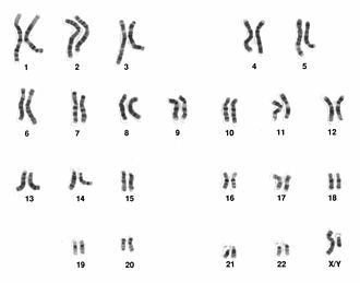
Human karyotype, methods of its examination
Human karyotype - set of 46 chromosomes (number – species specific characteristic)
- 22 homologous pairs of autosomes and 1 pair of sex chromosomes
- arranged and numbered according to the chromosome size and type (centromere position) and banding pattern into homologous pairs and seven chromosome groups
- Group A (1-3) – large metacentric chromosomes (but chr. 2 – submetacentric)
- Group B (4-5) – large submetacentric
- Group C (6-12, X) – medium-sized submetacentric
- Group D (13-15) – medium-sized acrocentric
- Group E (16-18) – short submetacentric
- Group F (19-20) – short metacentric
- Group G (21-22, Y) – short acrocentric (but chr. Y – submetacentric)
Karyotype analysis[edit | edit source]
Karyotype analysis (karyotyping) – analysis of banded chromosomes in light microscope; chromosomes are visible only in condensed form during cell division – cell cultivation is necessary.
Samples: in general – nuclear and dividing cells (e.g. erytrocytes or neurons are excluded); in practise – routinely only some types of samples are used:
- prenatally – chorionic villi sampling (CVS), amniotic fluid, fetal blood (PUBS – percutaneus umbilical blood sampling), tissues of spontaneously aborted fetus;
- postnatally – peripheral blood (lymphocytes), bone marrow, fibroblasts.
Cultivation – incubator (37 ºC, culture medium, stimulation of growth), 72 hours (blood samples), longer – approx. 2 weeks (prenatal, tissue cultures).
Colchicine – stops the cell division, interacts with the spindle, cells are arested in metaphase (c-metaphase) Cytogenetic processing: hypotonic saline, fixation, dropping of the cell suspension onto the slide, staining of preparations.
Types of staining[edit | edit source]
Conventional – only Giemsa, monochromatic chromosomes
Differential staining – banding
- G-banding: trypsin digestion + Giemsa, dark and light bands (dark bands – heterochromatin, gene-poor, adenine-thymine (AT) rich usually repetitive sequences; light bands – euchromatin, gene-rich, guanine-cytosine (GC) rich, unique sequences)
- Q-banding: historically first banding; quinacrine – non-stable fluorescent dye, the same banding pattern as G-bands
- R-banding: reverse banding pattern to G-bands; alkaline reagents and high temperature treatment
Selective staining
- C-banding: staining after denaturation in alkaline solutions; selectively highlights centromeres and another constitutive heterochromatin regions (namely in chromosomes 1, 9, 16 and Y)
- Ag-NOR banding (silver staining): selectively stains nucleolar organizing regions (NORs) located on the satellite stalks of the acrocentric chromosomes


