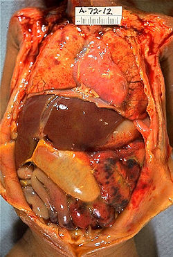
Infections in the neonatal period
Among infections in the newborn age we include:
- neonatal sepsis;
- meningitis;
- conjunctivitis;
- necrotizing enterocolitis;
- omphalitis;
- osteomyelitis, arthritis;
- mastitis;
- paronychium.[1]
Prenatal infections are also manifested in the newborn age:
Risk Factors[edit | edit source]
Fetal exposure to infection (acute infection of the mother, colonization of the birth canal) and immature immunity of the child (absence of IgM, low level of complementu). Furthermore, prematurity low birth weight, premature outflow of amniotic fluid, infection of the mother around birth (urinary tract infection), cloudy amniotic fluid (chorioamnionitis), poor social conditions of the mother , children with immunity defects and metabolic defects.[1]
The gateway of infection is usually the ascending route during the outflow of amniotic fluid (inhalation, smearing of the mucous membrane, conjunctiva) and the umbilical cord.
Most common agent: Streptococcus agalactiae (GBS), Enterobacter, Chlamydia, Mycoplasma, Varicella-zoster virus (VZV), Herpes simplex virus (HSV), Enteroviruses.
Postnatal Sepsis[edit | edit source]
Neonatal sepsis is a bacterial disease characterized by clinical symptoms with bacteremia. In 25% of cases, it also affects the meninges (meningitis) and contributes significantly to the morbidity and mortality of newborns. It affects approximately 2% of live births (incidence increases with immaturity), with premature outflow of amniotic fluid (PROM) the incidence is 3−5%.[2]
- Early sepsis
- By 3rd day, high mortality, fulminant course.
- Agents: most often GBS and Escherichia coli, also Staphylococcus aureus, Streptococcus pneumoniae and Listeria .
- Late sepsis
- After the 3rd day, often during a stay in the ICU. More often they are localized (pneumonia, meningitis, pyelonephritis).
- Agents: mainly G− (E. coli, Klebsiella, Pseudomonas), but also S. aureus and S. epidermidis, Serratia, Candida albicans, Listeria, Haemophilus influenzae.
Risk factors for the development of infection are immaturity, maternal infection, premature outflow of amniotic fluid (PROM), adnate infection, conditions after CPR, asphyxia, meconium aspiration, invasive procedures. The main vector of infection transmission is the hands of staff. Infection can penetrate hematogenously, transplacentally, during aspiration of infected amniotic fluid, colonization of the skin or intestine of a newborn, vertical transmission from mother to newborn during childbirth.[2]
Clinical manifestations
- Non-specific, variable - behavioral changes, thermostability, tachypnea, tachycardia, hypotension, poor peripheral circulation, skin manifestations (pallor, icterus, bleeding manifestations), anorexia/vomiting/diarrhea, metabolic symptoms.
- Therapy
- Initially empirical (until culture and sensitivity results), we use two-combination ATB' (ampicillin + cefotaxime). For this, we provide supportive therapy to ensure vital functions (complex intensive care, monitoring) and stabilize the internal environment.
- For early sepsis – aminopenicillin' + aminoglycoside 7–10 days i. v. (Augmentin + Gentamicin).
- For late sepsis - we try to target according to the population of the department, or broad spectrum ATB.
Meningitis[edit | edit source]
Inflammation of the meninges due to perinatal bacterial, rarely viral (HSV) infection. Most often in the 1st month of life. High mortality (30-60%), over 50% of survivors have permanent consequences. Immaturity or deficiency of cellular immunity and phagocytosis (mainly premature babies) is important for the development of the disease.
The most common agents are G− bacteria (E. coli, Klebsiella), then G+ cocci (streptococci B (GBS), rarely S. aureus), Listeria and from HSV-2 viruses. Clinical signs with early onset are similar to neonatal sepsis. The late form (>48 hours after birth) is characterized by: irritability, convulsions, arching of the fontanelle, ophthalmoplegia, hemiparesis, cranial nerve paresis and meningeal phenomena. With a viral etiology, pallor, respiratory insufficiency, fever, icterus and hepatic lesions are also present.
The result of neonatal meningitis can be a deficit of intellect, motor functions, sensory disorders, epilepsy, microcephaly, and mild behavioral disorders.
The drug of first choice is the double combination ATB ampicillin + gentamicin, further according to sensitivity (e.g. third-generation cephalosporins (ceftriaxone, cefotaxime) are suitable), in case of viral etiology - aciclovir. We also provide basic vital functions (crystalloids, adjustment of homeostasis, fluid balance, ventilation support, nutrition). As a precautionary measure in case of known maternal infection, caesarean section is performed.
Conjunctivitis[edit | edit source]
The most common agents are chlamydia, staphylococci, streptococci, Haemophilus influenzae, E. coli. As a preventive measure, we can already drip the eyes with Ophthalmo-Septonex in the delivery room. Therapy for mucus secretion – Ophthalmo-Septonex, for suppuration we administer ATB (Pamycon, Floxal) locally. In the case of long-lasting secretion unresponsive to ATB, it is necessary to think about the obstruction of the lacrimal ducts. If purulent conjunctivitis persists, poorly responding to local ATB, it is necessary to think about "Chlamydia trachomatis" (use overall macrolides).
Necrotizing enterocolitis[edit | edit source]
Hemorrhagic-necrotic inflammation of the intestine, which arises as a result of a disorder in the adaptation of the digestive system to food intake. Relatively common, it tends to be present in premature newborns. The cause of inflammation are G- rods (Pseudomonas, Klebsiella) and clostridia. It arises during the introduction of enteral nutrition. The source of infection is usually the mother or nursing staff. It usually starts 3-7 days after enteral nutrition is started.
May resemble neonatal sepsis (deterioration). It is manifested by abdominal distention, vomiting and blood in the stool. The prognosis is serious, fatal in 10-30%. Bowel perforation and peritonitis can be complications.
For treatment, we choose ampicillin + aminoglycoside (gentamycin) + clindamycin. Next, we discontinue enteral nutrition, which we replace with parenteral nutrition. We perform bowel decompression using NGS.
Other infections[edit | edit source]
- Omphalitis
- Manifests as redness and purulent secretion from the navel, with dissemination there are also signs of general infection. The most common agent is S. aureus. During treatment, we use ATB according to sensitivity. First-line drugs are first-generation cephalosporins, penicillin stable to beta-lactamases or ampicillin. Furthermore, it is necessary to clean the base of the navel and hospitalize the newborn.
Mastitis
- Most often in 2.-3. week. Clinically, there is redness, swelling, pain and purulent secretion of the mammary gland. We treat ATB according to sensitivity. In the case of an abscess, we choose incision and drainage.
- Diff. dg.: hormonal reaction (Halban's reaction) must be distinguished.
Osteomyelitis, arthritis
- The proximal humerus and femur are most often affected. Limitation of limb mobility, soreness, redness or swelling, possibly general symptoms. Erythrocyte sedimentation rate is typically high, x-ray may be normal initially.
- Paronychium
- Prevention is not to cut nails in the first weeks of life. In case of isolated occurrence, treat locally (wipe with alcohol, baths in hypermanganese, Framykoin). In the case of multiple occurrences of ATB in general – the drug of first choice is cephalosporins of the first generation.
Links[edit | edit source]
Related Articles[edit | edit source]
External links[edit | edit source]
- JEŽOVÁ, Marta – HOTÁRKOVÁ, Sylva – MŮČKOVÁ, Katarína. Hypertextový atlas novorozenecké patologie : Multimediální podpora výuky klinických a zdravotnických oborů [online]. Portál Lékařské fakulty Masarykovy univerzity [online], ©2010. [cit. 15.2.2023]. <http://portal.med.muni.cz/clanek-527-hypertextovy-atlas-novorozenecke-patologie.html>.
References[edit | edit source]
Source[edit | edit source]
- BENEŠ, Jiří. jirben.wz.cz : Studijní materiály [online]. [cit. 15.2.2023]. <http://jirben.wz.cz>.
References[edit | edit source]
- HRODEK, Otto – VAVŘINEC, Jan – A KOLEKTIV,. Pediatrie. 1. edition. Praha : Galén, 2002. ISBN 80-7262-178-5.
- ROZSYPAL, Hanuš. Základy infekčního lékařství. 1. edition. Praha : Karolinum, 2015. pp. 566. ISBN 978-80-246-2932-2.


