Intrinsic and efferent connections of cerebellum
The cerebellum is located in the posterior cranial fossa , dorsal to the medulla oblongata and the pontine ( brainstem ).
Rounded dorsally arched formation. Round, longitudinal, narrow central band, separated by sagittal depressions from the lateral parts = vermis cerebelli (cerebellar worm). Hemisphaeria cerebelli : 2 lateral, larger, symmetrically arranged hemispheres. Cranial surface flatter, in contact with the roof-like duplication of the dura mater ( tentorium cerebelli ). Dorsal and caudal surfaces arched; located in the fossae of the occipital bone under the transverse arms of the eminentia cruciformis (fossae occipitales cerebellares). The falx cerebelli (from the crista occipitalis interna) extends between the hemispheres of the cerebellum.
From the brainstem, 3 pairs of peduncles, pedunculi cerebellares, enter the cerebellum :
- Inferiores (corpora restiformia) – connect the oblongata with the cerebellum; line the caudal part of the rhomboid fossa.
- Medial (pontine; brachia pontis) – connect the pons with the cerebellum; border the rhomboid fossa.
- Superiores (brachia conjunctiva) – connect the tegmentum mesencephali with the cerebellum; border the rostral part of the fossa rhomboidea.
All peduncles contain pathways going to and from the cerebellum. Between the superior cerebellar peduncles is the velum medullare superius (craniale) – the anterior part of the roof of the fourth ventricle , drawn up to a peak called the fastigium.
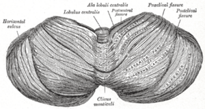
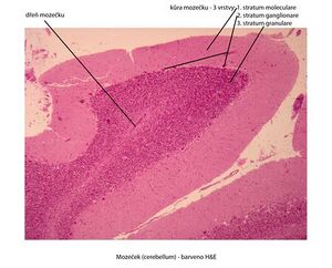
On the surface of the cerebellum, numerous transverse grooves separate the individual sections on the vermis and hemispheres = fissurae cerebelli . The largest and deepest fissures separate the 3 main sections: lobi cerebelli . Smaller fissures further divide these lobes into lobules: symmetrically located on the hemispheres; they correspond to the unpaired section on the vermis. The smallest fissures separate parallel strips of the cerebellum surface = folia cerebelli . The surface is covered with continuous gray matter: cortex cerebelli . Fissura prima - from the center to both sides, fissura horizontalis - dorsal pole, fissura posterolateralis - separates the nodulus and flocculus.
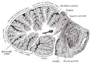
Inside the cerebellum is the white matter, corpus medullare . It projects in the form of discs as laminae albae into the folia of the cerebellum. On a sagittal section, the vermis forms a tree-like pattern ( arbor vitae , tree of life).
Paired clusters of gray matter are located in the white matter: cerebellar nuclei :
- Ncl. dentatus – the largest of the cerebellar nuclei. Two parts: dorsomedial (paleocerebellar) with fibers going to ncl. ruber and ventromedial (neocerebellar) with fibers going to the thalamus. The appearance of a wrinkled sac with an opening ventromedial to the mesencephalon . The opening of the sac = hilum (hilus) nuclei dentati. From here the path contained in the pedunculus cerebellaris superior.
- Ncl. emboliformis – elongated small nucleus, shaped like a blood clot. Located sagittally at the hilum of ncl. dentatus.
- Ncl. globosus – paired, located medial to ncl. emboliformis. Consists of several small spherical formations of gray matter.
- Ncl. fastigii – paired, located most medially at the fastigium, close to the midline.
Ncll. emboliformis, globosus and fastigii efferent to ncl. ruber, reticular nuclei, mesencephalon, pontine and oblongata. All cerebellar nuclei are the starting point of pathways emerging from the cerebellum – through them the cerebellum is involved in the movement control system
The nuclei contain the bodies of multipolar neurons, on which the axons of Purkinje cells terminate.
Morphological division of the cerebellum[edit | edit source]
It is divided by transverse grooves into 3 lobes . In each lobe, lobules are separated by smaller grooves (on the vermis and hemispheres). Morphological division allows for topographic orientation on the cerebellum, but does not correspond to the developmental and functional division.
Description of formations [ edit | edit source ][edit | edit source]
Upper surface (from anterior to posterior)
Vermis (lobus cerebelli anterior)
- Lingula cerebelli – 1 to several folia resting on the velum medulare superius.
- Lobulus centralis – a quadrangular group of foils in the anterior incisura. (fissula precentralis)
- Monticulus – the larger part of the upper surface of the vermis, convex in a hillock shape; it is divided into the culmen and the declivity (fissura prima) by a transverse groove
- Folium vermis – single leaf in the posterior cerebellar notch
Hemispheres
- Vinculum lingulae cerebelli – narrow white band.
- Ala lobuli centrales – a triangular group of folia in the anterior incisura.
- Lobulus quadrangularis – divided by a transverse groove into pars sup. et inf. (pars inf. otherwise also lobulus simplex).
- Lobulus semilunaris sup. – crescent-shaped.
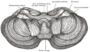
Lower surface (separated from the upper surface by the horizontal cerebellar fissure)
Vermis (posterior cerebellar lobe)
- Tuber vermis – protrudes into the posterior cerebellar fissure (fissura horizontalis)
- Pyramis vermis – the widest part of the worm (fissura prepyramidalis)
- Uvula vermis – an elongated elevation of several folia (fissura secunda)
- Nodulus vermis – attached to the uvula, rests on the velum medullare inf.
Hemispheres
- Lobulus semilunaris inf – it is joined by lobulus gracilis.
- Lobulus biventer – convex.
- Cerebral tonsils – significantly convex groups of horseshoe-shaped foci.
- Flocculus – a stalked group of folia with a curly margin; a rudimentary paraflocculus is attached at the posterior margin.
Divisions of cerebellum according to developmental relationships[edit | edit source]
Vestibular cerebellum – the oldest part, the basis of development is the vestibular pathways. It consists of: flocculus, lingula and nodulus.
Spinal cerebellum – the basis of development is the spinocerebellar pathways, it divides the vestibular cerebellum into the anterior (lingua) and posterior (nodulus and flocculus) parts. It consists of the anterior: lobulus centralis, culmen, lobulus quadrangularis superior and the posterior: pyramis, uvula, paraflocculus.
Cerebral cerebellum – develops by afferentation from the cortex, through the pontocerebellar tract. It arises in the middle of the older parts of the cerebellum collectively referred to as the palaeocerebellum. It is itself then called the neocerebellum (seu lobus medius).
The striation of the cerebellum occurs gradually.
The sulcus primarius (fissura prima) is formed earliest. It separates the part called the anterior (rostral) lobe. This includes the lingua and the anterior part of the spinal cerebellum. The following groove - fissura praepyramidalis delimits the middle lobe and the caudalis lobe in the vermis region. Fissura nodulouvularis - the boundary between the caudalis lobe and the pars nodulofloccularis.
Division involving both anatomical and developmental characteristics[edit | edit source]
- Lobus rostralis (anterior) – anterior rudiment of the vestibular cerebellum and anterior spinal cerebellum.
- Lobus medius – the largest part, includes the cerebral cerebellum.
- Lobus caudalis (posterior) – posterior spinal cerebellum.
- Lobus nodulofloccularis – part of the vestibular cerebellum not included in the lobus rostralis.
Functional connectivity of the cerebellum[edit | edit source]
Afferent pathways via the inferior, medial, and superior cerebellar peduncles to the cerebellar cortex. From the cerebellar cortex, fibers exit and end in the cerebellar nuclei.
The cerebellar nuclei send axons to the gray matter of the trunk (mainly to the reticular formation , ncl. ruber , to the thalamus ). From the gray matter of the trunk, pathways to the spinal cord , which influence cells that send their axons as motor fibers to skeletal muscles . It controls and controls motor activities and muscle tone, with the vermis participating in the coordination of the trunk muscles and the hemisphere of the ipsilateral limb muscles.
Cerebellar pathways[edit | edit source]
Afferent pathways are directed mainly to the cerebellar cortex. Efferentation begins with Purkinje cells (1st neuron) and continues after switching in the cerebellar nuclei (2nd neuron) into centrifugal pathways going outside the cerebellum.
Vestibulocerebellum - lobus flocculonodularis and lingula vermis are connected to the vestibular nuclei of the rhombencephalon, from the ncc. vestibulares and ncc. colliculi superiores and the visual cortex, maintaining body balance, spatial orientation.
Spinocerebellum - posterior and anterior cerebellar lobes, except for the rostral section of the lingula, it connects to the spinal cord, af. fibers go from the trigeminal nerve, auditory and visual structures of the CNS and ef. fibers go to the motor nuclei of the brainstem, its function is to coordinate motor activity and respond to proprioceptive information.
Cerebrocerebellum - through the nuclei pontis connected to the cerebral cortex, af. fibers from the cerebral cortex through the ncc. pontis, ef. are to the motor thalamus ( nc. ventralis anterior and lateralis ) and to the cerebral cortex, the function is coordination and timing of muscle movements.
A) Pathways of the nodulo-floccular part:
- Afferentation from the vestibular nerve as the vestibulocerebellar nerve.
- The efferentation goes to Deiters' nucleus, according to its origin as tr. nodulovestibularis and flocculovestibularis. From the ncl. fastigii, the tr. fastigiovestibularis (Russell's bundle) goes more independently along the pedunculus cerebellaris inf. to the Deiters' nucleus.
B) Pathways of the lobi rostralis and lobi caudalis Afferent pathways:
- Tr. spinocerebellaris posterior.: Stilling-Clark nucleus - pedunculus cerebellaris inferior. - cerebellar cortex (and cerebellar nuclei).
- Tr. spinicerebellaris ant. (Gowersi) : crossing in the spinal cord - lateral spinal cord (ventrally from tr. spinocerebelaris post) - through the pedunculi cerebellares superiores into the cerebellum.
- Tr. bulbocerebellar : from the nuclei of the posterior spinal cord - uncrossed as dorsal external arcuate fibers or crossed as ventral external arcuate fibers and internal arcuate fibers - via the inf. cerebellar peduncle.
- Tr. nucleocerebellares : from the nuclei of the sensitive cranial nerves.
- Tr. olivocerebellares : from the main olive (older part) and the secondary olive.
- Tr. tectocerebellaris : from the gray matter under the superior colliculi - velum medullare superior - cortex vermis superior.
- Tr. reticulocerebellaris : from the nuclei of the lateral nuclei RF - pedunculus cerebelaris inf. - vermis - to the ipsilateral hemisphere.
- Tr. rubrocerebellaris : after crossing tr. rubrospinalis it branches into - pedunculus cerebellaris sup.
Efferent pathways:
- Tr. cerebellotegmentalis (dentatotegmentalis) : from cerebellar nuclei (mainly ncl. dentatus) - pedunculus cerebellaris sup. - nuclei of FR pontine and mesencephalon.
- Tr. cerebellorubralis (dentatorubralis) : via the pedunculi cerebelli sup. to the ncl. ruber (further via the tr. rubrospinalis and rubroolivaris to the olive).
- Tr. cerebelloolivaris : through the pedunculi cerebelli inf. - contralaterally to the main olive (older part) and the secondary olive.
- Tr. cerebellotectalis : via the pedunculi cerebelli sup.
- Tr. cerebellothalamicus (embolothalamicus): through the central nuclei of the thalamus to the striatum.
C) Pathways of the medial lobe
Afferent pathways:
- Tr. pontocerebellares : from ncll. pontis, where the - pedunculi cerebelli medii - cerebellar cortex cross; the pathway is a continuation of the corticopontine pathway (tr. frontopontinus et tr. occipitotemporopontinus) and connects the hemispheres of the forebrain and cerebellum in a cross-like manner.
- Tr. olivocerebellares : as in the lobi rostralis et caudalis pathways, but originates from the neooliva (younger part of the olivary nucleus).
- Tr. corticocerebellares : from the motor area of the frontal lobe - pedunculi cerebelli inf. - to the ipsilateral hemisphere of the cerebellum.
Efferent pathways:
- Tr. cerebellorubrales : conduction as in the path of the same name from the previous group of paths.
- Tr. cerebrothalamici : from ncl. dentatus to ventrolateral nuclei of the thalamus (hereinafter referred to as tr. thalamocorticalis to areas 4 and 6).
Links[edit | edit source]
Related articles[edit | edit source]
External links[edit | edit source]
- JANČÁLEK, Radim – DUBOVÝ, Petr. Základy neurověd v zubním lékařství [online]. MEFANET, ©2011. The last revision 27.10.2011, [cit. 26.11.2011]. <http://portal.med.muni.cz/clanek-560-zaklady-neuroved-v-zubnim-lekarstvi.html>.
- DUBOVÝ, Petr. Základy neuroanatomie a nervových drah : Multimediální podpora výuky klinických a zdravotnických oborů [online]. Portál Lékařské fakulty Masarykovy univerzity [online], ©2007. The last revision 3.6.2011, [cit. 2011-11-27]. <http://portal.med.muni.cz/clanek-442-zaklady-neuroanatomie-a-nervovych-drah.html>.
Reference[edit | edit source]
Source[edit | edit source]
- BOROVANSKÝ, Ladislav. Soustavná anatomie člověka. 3. přeprac. vyd. Praha: Státní zdravotnické nakladatelství, 1967, S. 597-1023.


