Joints of the upper limb
From WikiLectures
HK joints include the connection of the HK girdle with the sternoclavicular (articulatio sternoclavicularis) and acromioclavicular (articulatio acromioclavicularis), free HK joints are the shoulder joint (articulatio humeri), the elbow joint (articulatio cubiti), the distal radioulnar joint (articulatio radioulnaris distalis) and the joints of the hand (articulationes manus).
HK girdle junction (Juncturae cinguli membri superioris)[edit | edit source]
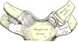
Articulatio sternoclavicularis[edit | edit source]
- it is a compound joint where two bones meet - it connects the clavicle to the manubrium sterni
- head: extremitas sternalis claviculae , socket: incisura clavicularis sterni
- the sheath is short and stiff
- the joint contains the discus articularis (from fibrocartilage) – compensates for the uneven curvature of the joint surfaces, the strengthening is noticeable cranially and dorsally, connected to the joint capsule around the entire circumference
- sheath reinforced by ligaments
- ligamentum sternoclaviculare anterius et posterius – attached to the capsule in front and behind
- ligamentum interclaviculare – connects both collarbones (clavicles)
- ligamentum costoclaviculare – ensures the connection of the clavicle to the 1st rib
- movements in all directions on a small scale
- with sharp impacts transferred from HK, a fracture of the clavicle occurs rather than a dislocation of the joint due to the strength of the capsule and ligaments
- vascular supply: a. thoracica interna, sometimes also a. thoracica superior
- nerves: they come from the nn. supraclaviculares mediales, also n. subclavius
Articulatio acromioclavicularis[edit | edit source]
- flat joint (possible presence of discus articularis), connects the outer end of the clavicle with the acromium
- the articular surfaces are flat, formed by facies articularis acromii and extremitas acromialis claviculae
- the capsule is short and stiff, with a thickening cranially
- ligamentum acromioclaviculare – strengthens the upper side of the joint capsule
- the movements of this joint are small, supplementary movements are due to the sternoclavicular joint, there may be a small discus articularis
- the movements between the clavicle and the scapula are directed by the coracoclavicular ligament , which connects the processus coracoideus with the lower surface of the clavicle (this ligament limits the movements of the scapula) - it consists of 2 components: the ligamentum trapezoideum and the ligamentum conoideum
- ligamentum coracoacromiale - connects the processus coracoideus with the acromium (it is a strong ligament stretched over the shoulder joint), the older name is fornix humeri
- ligamentum transversum scapulae superius - complements the scapula incisura for the suprascapularis nerve (+ ligamentum transversum scapulae inferius)
Joints free HK (Articulationes membri superioris liberi)[edit | edit source]
Articulatio humeri[edit | edit source]
- the geometric type is a spherical free joint
- articular surfaces consist of: caput humeri , cavity: cavitas glenoidalis , which is expanded by the cartilaginous edge of the labrum glenoidale
- the articular capsule attaches to the scapula at the outer circumference of the labrum glenoidale, goes around the circumference of the collum anatomicum humeri, but descends medially to the collum chirurgicum and folds into the folds.
- the strengthening of the joint capsule is with the help of tendons around the walking muscles and also joint ligaments
- rotator cuff = group of muscles and tendons strengthening the joint capsule (4 muscles):
- m. supraspinatus
- m. infraspinatus
- m. teres minor
- m. subscapularis (between the tendon of this muscle and the capsule there is a bursa subtendinea musculi subscapularis)
- Ligaments:
- ligamentum coracohumerale – in front
- glenohumeral ligaments – there are 3, located in the front of the capsule
- ligamentum coracoacromiale – above the joint
+ the tendon of the biceps brachii muscle is clamped in this place (from tuberculum supraglenoidale to sulcus intertubercularis)
- Bursae mucosae: (are formed in places of pressure and friction)
- bursa subtendinea musculi subscapularis
- subcoracoid bursa
- bursa subdeltoidea - often irritated when carrying objects on the shoulders - development of bursitis
- Shoulder joint movements: (wide range, all directions, it is the most mobile joint)
- ventral flexion - forearm flexion (80°)
- dorsal flexion (extension) - grip
- abduction – abduction, only to the horizontal (then it blocks the coracoacromial ligament)
- adduction - bearing
- circumduction – combination of previous movements
- middle position - slight abduction and flexion
- due to the small articular fossa compared to the head, dislocation is a common injury
- anterior dislocation – in the vast majority of cases, a so-called Bankart defect occurs when the glenoid labrum breaks
- arteries: the strongest ascending branch in front, following the tendon of the biceps brachii muscle, is referred to as Laing's artery
- nerves: the nerves of the shoulder joint come from the suprascapularis nerve, from the nn. subscapulares and n. axillaris
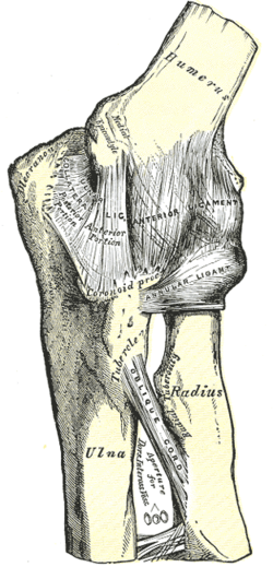
Articulatio cubiti[edit | edit source]
- it is a compound joint because 3 bones meet there (humerus, radius and ulna)
Art. humeroulnaris[edit | edit source]
- the pulley joint that is between the trochlea humeri and incisura trochlearis ulnae
Art. humeroradialis[edit | edit source]
- the ball-and-socket joint that is between the capitulum humeri and the fovea capitis radii
Art. radioulnaris proximalis[edit | edit source]
- the wheel joint, which is between the incisura radialis ulnae and the circumferential articularis caput radii
- the articular capsule surrounds all 3 joints, leaving the epicondyles free due to the forearm muscles - on the ulna, it attaches along the edge of the articular surfaces and on the radius, it descends to the neck as a recessus sacciformis
- Ligaments:
- ligamentum collaterale radiale et ulnare – along the edges
- ligamentum annulare radii – has its beginning and attachment on the ulna, wraps around the collum radii (ringbone), has 3 stripes:
- ligamentum olecranohumerale - between the olecranon and the epicondyle
- ligamentum humerocoronoideum - between the epicondyle and processus coronoideus
- ligamentum obliquum - between the olecranon and the coronoid process
- Bursae = weight pouches:
- bursa subcutaneous olecrani - on the olecranon, between the attachment tendon of the triceps brachii muscle
- bursa subtendinea musculi tricipitis brachii
- bursa bicipitoradialis - between the tendon of the triceps brachii muscle and the front surface of the tuberostias radii
- Moves:
- the base position is extension
- medium stance – slight flexion, slight pronation
- flexion and extension – in the humeroulnar joint and simultaneously in the humeroradial joint, in the range of 125–145 °, the extension is stopped by the olecranon resting on the fossa olecrani, in the case of a smaller olecranon, hyperextension is possible (physiologically in women)
- pronation and supination – ensure humeroradial, radioulnar proximal joints and accompanied by movement of the radioulnar distal joint (see below)
- musculi articulares = muscle bundle that prevents the capsule from twisting during movements
- membrana interossea antebrachii – fibrous membrane between the margins of the interossei of the radius and ulna , the fibrous bundles run diagonally from the radius distally to the ulna, in the proximal part there is a chorda obliqua – a strip of bundles running in the opposite direction
Art. radioulnaris distalis[edit | edit source]
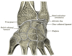
- cylindrical joint, it is the articulation of the distal ends of the radius and ulna (caput ulnae and incisura ulnaris radii)
- the case is free and allows combined movements with art. humeroradialis and art. radioulnaris proximalis – pronation and supination
- Moves:
- pronation – the ulna is in the same position, proximally the radius rotates around its long axis, distally around the head of the ulna
- supination – the basic position of the joint, bones next to each other, palm ventrally
- Arteries: come from the anterior interossea artery and from the posterior interossea artery
- Nerves: there are branches from the anterior interosseus nerve and the posterior interosseus nerve
Articulationes manus – joints of the hand[edit | edit source]
Art. radiocarpalis[edit | edit source]
- compound elliptical joint
- head: os scaphoideum , os lunatum , os triquetrum , fossa: facies artilcularis carpalis radii , ulna does not participate in articulation thanks to the discus articularis – ulnarly connects to the fossa
- the sleeve is loose, allowing a wide range of movement
- Moves:
- palmar flexion and extension – 150–170 °
- ulnar – 35 ° and radial duction – 15 °
- the combination creates circumduction
- basic position – continuation of the long axis of the forearm
- middle position - as basic
- movements combined into a functional unit with art. mediocarpalis, art. carpometacarpales, intercarpales, intermetacarpales (see below)
Art. mediocarpalis[edit | edit source]
- compound joint
- head medially: os capitatum , os hamatum , fossa medially: os scaphoideum , os lunate , os triquetrum , head radially: os scaphoideum , fossa radially: os trapezium , os trapezoideum
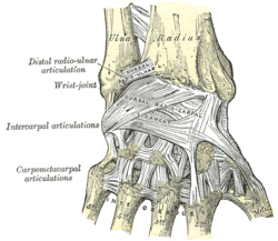
Articulationes intercarpales[edit | edit source]
- articulation of the lateral surfaces of the carpal bones with each other
- strengthening ligaments: ligamenta intercarpalia palmaria, dorsalia et interossea
Articulationes carpometacarpales[edit | edit source]
- they connect the distal row of carpal bones to the bases of the metacarpal bones
Art. carpometacarpalis pollicis[edit | edit source]
- saddle joint
- head: base of the 1st metacarpal , socket: trapezium axis
- Moves:
- palmar
- dorsiflexion
- abduction
- adduction
- slight rotation, thumb opposition
Ligaments of the hand[edit | edit source]
- ligamenta collateralia carpi – runs along the sides of the wrist
- ligamentum ulnocarpale palmare, lig. ulnocarpal dorsale, lig. radiocarpale palmare et dorsale – ligaments extend from the radius and ulna across the middle of the carpus, obliquely and distally to the opposite edge
- ligamentum carpi radiatum – palmarly from the os capitatum in all directions to the surrounding bones
- ligamenta intercarpalia interossea, ligamenta metacarpalia interossea
- between the eminentia carpi radialis et ulnaris the retinaculum musculorum flexorum is opened , its connection with the carpus creates the canalis carpi – here the course of the flexor carpi radialis, flexor digitorum superficialis et profundus, flexor pollicis longus, median nerve
- Carpal tunnel syndrome - caused by permanent compression of the median nerve, which subsequently causes problems such as tingling, tingling and burning, loss of sensitivity, stiffness
Articulationes metacarpophalangeae[edit | edit source]
- palmarly cylindrical, distally spherical surface of head
- head: metacarpal bones , socket: proximal phalanges
- cases free
- Ligaments:
- ligamenta collateralia – on the sides
- ligamenta palmaria – on the palmar side, including the fibrous plate: fibrocartilago palmaris – strengthens the sheath, the fibrous layers of the tendon sheaths of the flexors are attached to the plates
- lig.metacarpale transversum profundum - connect the cases of adjacent metacarpoph.joints
- Moves:
- flexion and extension
- abduction and adduction
- rotation
- circumduction - only if the articulation is in a position where the spherical part of the head is, adduction and abduction is not possible when the finger is flexed and transferred to the cylindrical surface of the head
- basic position – outstretched finger
- middle position – with medium flexion
Articulationes interphalangeae[edit | edit source]
- pulley joints
- head: proximal and middle links , fossa: base of middle and distal links
- Ligaments:
- palmar ligament"
- "collateral ligament"
- "fibrocartilagines palmares
- Moves:
- flexion
- basic position: with the finger extended
- middle position: middle flexion
Links[edit | edit source]
Related Articles[edit | edit source]
- Articulatio humeri
- Articulatio cubiti
- Joints
- Hand joints
References[edit | edit source]
- ↑ GRIM, Miloš and Rastislav DRUGA, et al. Fundamentals of Anatomy 1. 1st ed. Prague: Galén, 2001. 159 pp. ISBN 80-7262-112-2 .
References[edit | edit source]
- CIHÁK, Radomír and Miloš GRIM. Anatomy. 2., edit. and add edition. Prague: Grada Publishing, 2002. 470 pp. Vol. 2. ISBN 80-7169-970-5 .
- GRIM, Miloš and Rastislav DRUGA, et al. Fundamentals of Anatomy 1. 1st ed. Prague: Galén, 2001. 159 pp. ISBN 80-7262-112-2 .


