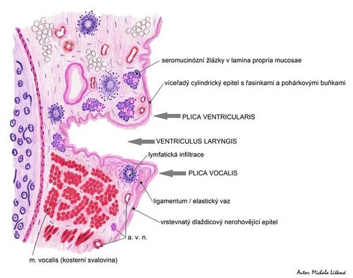
Larynx (histological slice)
From WikiLectures
On the frontal section of the larynx, we see multiple types of tissue such as muscle, various epitheliums, glands and cartilage.
Basic formations on the specimen that must be distinguished:
- Plica vestibularis:
- false vocal fold,
- multi-rowed cylindrical epithelium with cilia,
- lamina propria contains serous and mucinous glands,
- lymphatic tissue in places (dark purple accumulation of cells that forms "islets"),
- cranially connects to the laryngeal surface of the epiglottis.
- Plica vocalis:
- right vocal cord,
- stratified squamous epithelium without keratinization,
- the basis is Lig. vocale and striated muscle (M. vocalis), possibly (on a larger section) also M. thyroarytenoideus.
- Ventriculus laryngis:
- is a roughly diamond-shaped space between the two vocal cords.
The supporting tissue of the larynx consists of hyaline thyroid cartilage - Cartilago thyroidea (it is visible only on extensive sections). Cartilage can ossify.
Note: Stratified squamous epithelium without keratinization occurs in the airways in places that are forced to resist a higher pressure of air, or more friction.
Links[edit | edit source]
Related Articles[edit | edit source]
References[edit | edit source]
- MUDR. EIS, Václav – MUDR. JELÍNEK, – MUDR. ŠPAČEK, Martin. Histopatologický atlas [online]. ©2006. [cit. 2010]. <http://histologie.lf3.cuni.cz/histologie/atlas/index.htm>.
- JUNQUIERA, L. Carlos – CARNEIRO, José – KELLEY, Robert O.. Základy histologie. 1. edition. Jinočany : H & H 1997, 1997. 502 pp. ISBN 80-85787-37-7.


