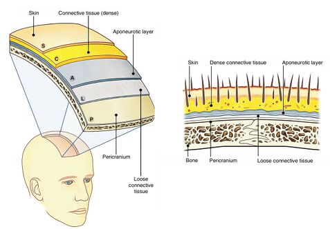
Layers of scalp, frontal and temporal regions
|
This article was marked by its author as Under construction, but the last edit is older than 30 days. If you want to edit this page, please try to contact its author first (you fill find him in the history). Watch the discussion as well. If the author will not continue in work, remove the template Last update: Sunday, 01 Dec 2024 at 10.36 pm. |
Layers of scalp, frontal and temporal regions[edit | edit source]
The scalp refers to the layers of skin and subcutaneous tissue that cover the bones of cranial vault. It extends from temporal fascia to the zygomatic arches.[edit | edit source]
The scalp consists of 5 layers. First 3 layers are tightly bound together and move as a collective structure. The scalp layers can be memorized using the mnemonics “SCALP”:
1. Skin- contains numerous hair follicles and sebaceous glands (thus a common site for sebaceous cysts).
2. Connective tissue (Dense subcutaneous)- connects the skin to the epicranial aponeurosis. It is richly vascularized and innervated. Blood vessels within the layer are highly adherent to the connective tissue. This enables the connective tissue to constrict fully and if so, the scalp can be a site of profuse bleeding.
3. Epicranial Aponeurosis (Galea Aponeurotica)- thin and tendon-like structure that connects the occipitalis and frontalis muscles.
4. Loose areolar connective tissue- thin connective tissue layer that separates the periosteum of the skull from the epicranial aponeurosis. It contains numerous blood vessels, including emissary veins which connect the veins of the scalp to the diploic veins and intracranial venous sinuses.
5. Pericranium- outer layer of the skull bones (periosteum of the skull). It becomes continuous with the endosteum at the suture lines.
Loose areolar connective tissue considered as the “danger area” of the scalp, since it contains the emissary veins- valveless veins which connect the extracranial veins of the scalp to the intracranial dural venous sinuses. The emissary veins are a potential pathway for the spread of infection from the scalp to the intracranial space.
Temporal and Frontal regions:[edit | edit source]
Temporal region include:[edit | edit source]
1. Temporal fossa
2. Temporalis muscle, originated from the anterior, posterior and superior borders of temporal fossa, inserted beneath the zygomatic arch. Innervated by mandibular nerve.
3. Temporoparietal fascia/ superficial temporal fascia has 2 layers that merge in the superior temporal line. Beneath the 2 layers there’s a fat pad.
4. Superior temporal artery, branch of external carotid artery. Courses above the superficial temporal fascia.
5. Auriculotemporal nerve, branch of the mandibular nerve.
6. Temporal branches of facial nerve.
7. Anterior and posterior branches of the deep temporal nerve.
8. Superior temporal vein accompanies the artery.
9. Temporal medial vein runs between the 2 layers of the fascia.
Temporal region borders:[edit | edit source]
1. Superiorly and posteriorly- superior temporal line
2. Inferiorly- zygomatic arch
3. Anteriorly - frontal process of the zygoma and zygomatic process of the frontal bone.
Temporal region layers:[edit | edit source]
The layers of the temporal region are different than the rest of the scalp.[edit | edit source]
1. Skin
2. Subcutaneous tissue
3. Superficial temporal fascia
4. Innominate fascia- contains the auriculotemporal nerve.
5. Deep temporal fascia
6. Musculature of the face.
7. Parotidmasseteric fascia of parotid gland and masseter muscle
8. Temporalis and Masseter muscles
9. Bones (Calvarium & basicranium)
Sources:[edit | edit source]
Stingl, J., Grim, M., & Druga, R. (2012). ''Regional anatomy''. Galen.


