Liver - structure, nutritional and portal vascular bed,intrahepatic bile ducts
From WikiLectures
Liver - Structure, Nutritional and Portal Vascular Bed, Intrahepatic Bile Ducts
Introduction-
The liver is the largest gland in the human body and plays vital roles in metabolism, detoxification, digestion, and bile production. It is located in the right upper quadrant of the abdomen and is intraperitoneal, except for the bare area.
Structure
Macroscopic Features:
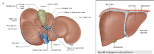
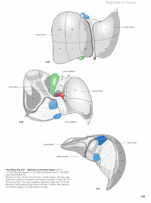
- Lobes:
- Right Lobe: Largest, accounting for approximately two-thirds of liver mass.
- Left Lobe: Smaller, separated from the right lobe by the falciform ligament.
- Caudate Lobe: Positioned near the inferior vena cava.
- Quadrate Lobe: Located near the gallbladder.
- Capsule:
- Enclosed by Glisson's capsule, a fibrous covering that extends into the liver along the vessels and ducts.
- Surface:
- Diaphragmatic Surface: Convex, facing the diaphragm.
- Visceral Surface: Concave, facing the abdominal organs.
- Ligaments:
- Falciform Ligament: Connects the liver to the anterior abdominal wall and diaphragm.
- Coronary and Triangular Ligaments: Attach the liver to the diaphragm.
- Ligamentum Teres: Remnant of the umbilical vein.
- Ligamentum Venosum: Remnant of the ductus venosus.
Microscopic Features:
- Lobules:
- Classical Lobule: Hexagonal structure centered around a central vein.
- Portal Lobule: Triangular structure with bile duct in the center.
- Acinus: Functional unit based on oxygen and nutrient gradients.
- Cell Types:
- Hepatocytes: Main functional cells performing metabolism and bile production.
- Kupffer Cells: Specialized macrophages within the sinusoids.
- Stellate (Ito) Cells: Store vitamin A and regulate extracellular matrix.
- Endothelial Cells: Line the sinusoids, fenestrated for exchange.
Nutritional and Portal Vascular Bed
Dual Blood Supply:
- Nutritional Supply:
- Hepatic artery (25% of total blood flow).
- Oxygen-rich blood from the celiac trunk.
- Portal Venous Supply:
- Portal vein (75% of total blood flow).
- Nutrient-rich, oxygen-poor blood from the gastrointestinal tract, pancreas, and spleen.
Vascular Pathway:
- Blood from the portal vein and hepatic artery mixes in the sinusoids.
- Flows centripetally toward the central vein.
- Drains into the hepatic veins and subsequently into the inferior vena cava.
Microcirculation:
- Sinusoids: Capillary-like vessels with fenestrated endothelial lining and no basement membrane, allowing exchange between blood and hepatocytes.
- Central Vein: Collects blood from the sinusoids.
Intrahepatic Bile Ducts
- Bile Canaliculi:
- Small intercellular channels formed by adjacent hepatocytes.
- Transport bile toward the portal triads.
- Canals of Hering:
- Small ducts connecting bile canaliculi to interlobular bile ducts.
- Lined by hepatocytes and cholangiocytes.
- Interlobular Bile Ducts:
- Located in the portal triads.
- Lined entirely by cholangiocytes.
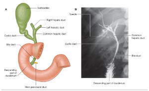
- Hepatic Ducts:
- Merge to form the right and left hepatic ducts.
- Join to form the common hepatic duct.
- Common Hepatic Duct:
- Joins the cystic duct from the gallbladder to form the common bile duct, which empties into the duodenum via the ampulla of Vater.
Diagrams
- Macroscopic Anatomy of the Liver:
- Labeled diagram showing lobes, surfaces, and ligaments.
- Classical Liver Lobule:
- Microscopic view illustrating the hexagonal structure, portal triads, sinusoids, and central vein.
- Bile Duct System:
- Flow of bile from canaliculi to the common bile duct.
- Dual Blood Supply:
- Schematic representation of the hepatic artery, portal vein, sinusoids, and venous drainage.
Sources
- Gray’s Anatomy for Students, 4th Edition.
- Sobotta Atlas of Human Anatomy, 16th Edition.


