Lymph node
A lymph node is a spherical or oval shaped organ (1 – 25mm). It is found in the circulation lymphatic vessels and serves as a biological filter of lymph. There are ~500 lymph nodes in the body. Lymph nodes can be found mainly in the armpit, in the groin, near the large vessels of the neck, in the chest or abdominal cavity. They play an essential role in the body's defense against microbes and tumor cells.They contain APC’s (lymphocytes, macrophages and dendritic cells).
Function: Filter lymph
Site of initiation of specific immune response: naïve lymphocytes circulate between lymph nodes searching for foreign antigen on APC’s 🡪 induce the activation, proliferation and differentiation of naïve lymphocytes into mature
Structure of the lymph node[edit | edit source]
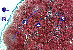
A simple lymph node is usually bean-shaped (kidney-shaped). Its size ranges from 1-25 mm. On the surface, it is covered by a capsule of dense collagenous tissue with elastic fibers and a small amount of smooth muscle cells. Fibrous septa, trabeculae, depart from this capsule and point to the center of the node. As with other peripheral lymphatic organs, the stroma of lymph nodes is reticular tissue. It consists of reticular cells, reticular fibers and fixed macrophages. Together, they form a three-dimensional network that is filled with free cells. The largest number of lymphocytes, macrophages are found here, we also find eosinophilic and basophilic granulocytes and plasma cells. fibroblasts may also be present in the fibrous bundles along the vessels. The lymph node consists of a peripherally located cortex (cortex) and a centrally located marrow (medulla). In the concavity of the node is the hilus. Nerves, arteries enter the lymph node at this point, and veins and usually one efferent lymphatic vessel leave it. Afferent lymphatic vessels enter the convexity of the node.
Cortex[edit | edit source]
The cortex contains oval-shaped lymphatic follicles filled with accumulated B-lymphocytes. It appears dark because the B-lymphocytes present here have a distinctly basophilic nucleus and condensed chromatin. In the follicles, we distinguish the cortical B-zone and the paracortical T-zone according to the amount of the respective B-lymphocytes or T-lymphocytes. Furthermore, there are follicular dendritic cells - reticular fibroblasts and macrophages, which have long processes, with the help of which they capture and present antigens to antigen presenting cells of the immune system. They are distinguishable from lymphocytes themselves by a lighter, oval-shaped nucleus. There are two types of follicles in the cortex:
- primary follicles - follicles with a dark appearance (the lighter central area is not indicated)
- secondary follicles - have visible bright germinal (germinative) centers that reflect the increased activity of the organism's defense - they are created by significant mitotic activity of lymphocytes. It contains activated B-lymphocytes (centroblasts, immunoblasts). These cells have finer chromatin and a larger volume of basophilic cytoplasm.
A darker zone separating the center from the paracortical zone is visible around the germinal center. Here there are abundant small B-lymphocytes that carry receptors for the given antigen. We call these B-lymphocytes cloned B-lymphocytes. They have a distinctly basophilic nucleus containing condensed chromatin.
The cortical zone is followed by a transition to the thymodependent paracortical T-zone, in which veins with high epithelium (HEV, high endothelial venules) run. Endothelial cells of these veins have a cubic shape and a light nucleus. The B-zone and the T-zone are difficult to distinguish from each other, so it is advisable to use immunohistochemical or histochemical methods.
Medulla[edit | edit source]
The medulla of a lymph node is the region between the paracortical zone and the hilum of the node. It is formed by anastomosing cords of lymphatic tissue, medullary sinuses are developed between them. There are fewer lymphocytes here than in the cortex, on the contrary, there are abundant macrophages with phagocytosed material. Furthermore, we find plasmocytes, sometimes also heparinocytes.
Hilum[edit | edit source]
On the concave surface of the node – contains afferent blood vessels and efferent lymph vessel.
Lymphatic sinuses and lymph circulation[edit | edit source]
Lymph is supplied to the node by afferent vessels (vasa afferentia) which enter on its convex surface. It continues through the cortex, medulla, reticular stroma and ligament to the hilum, from where it is removed by the draining lymphatic vessel (vas efferens). The draining lymphatic vessel is usually alone, or less often accompanied by another. The vessels have numerous valves that direct the flow of lymph. The entire node contains an abundance of lymphatic sinuses, which are spaces where lymph and B-lymphocytes flow. These sinuses do not have an endothelial lining, they are formed by littoral cells, which are reticular cells covered with reticular fibers. Pores run between them, the basement membrane is even missing. The function of the sinus system is to slow down the flow of lymph and thus enable its filtration. The thickness of the sinuses is not constant. This arrangement allows communication with the surrounding lymphatic tissue. The lymph proceeds first through the subcapsular sinuses (which are located in the cortex under the fibrous capsule). It then proceeds through the parafollicular sinuses and is fed into the medullary sinuses. From there it is carried away by efferent vessels.
Simplified: Lymph circulation: afferent lymph vessel 🡪 subcapsular sinus 🡪 capsular sinus 🡪 medullary sinus 🡪 efferent lymph vessel
Lymph nodule: primary (inactive)/ secondary (active) 🡪 3 zones:
- dark zone (centroblasts): rapidly proliferating B-lymphocytes
- light zone (centrocytes): the B-lymphocytes “compete” to bind to the unprocessed antigen, the unbonded B-lymphocytes will die.
- Mantle zone (quiescent cells): peripherally marginalized B-lymphocytes due to the rapid proliferation.
We therefore distinguish several types of sinuses depending on the position to the surrounding structures:
- sinus marginales/subcapsulares - under the fibrous capsule;
- sinus peri(inter-)folliculares - in the cortex between individual follicles;
- sinus paratrabeculares - in the cortex along the trabeculae;
- sinus medullares - in the medulla.
Types of lymph nodes:[edit | edit source]
- Tributary region: a specific area drained by a group of lymph nodes
- Regional lymph nodes: a group of lymph nodes draining a specific anatomic region
- Sentinel lymph node: the 1st node/group of nodes draining a cancer – the 1st lymph node/group to be invaded by mediatizing cancer cells (1st alarm) – swelling always indicates a pathological process
Non-encapsulates lymphatic tissue - MALT (GIT)[edit | edit source]
Lymphoid tissue located both diffusely and in nodules throughout
the body – forms mucosal immune system – MALT (mucosa-associated lymphoid tissue): contains the same number of lymphocytes as the rest of the body. MALT forms barriers against pathogens in the GIT and respiratory system, skin and other organs.
Types:
- D-MALT (diffuse): diffuse cells mainly in the GIT
- O-MALT (organized): concentration of lymphoid tissue:
- Solitary lymphoid nodules
- Aggregated lymphoid nodule
- Peyer’s patches: in the ileum
- Lymphoid nodules of the vermiform appendix
Other places where Lymphatic Tisuues are present are
- nose
- Bronchus
- Skin : keratinocytes, mast cells, Langerhans cells
- conjunctiva
- urninary tract
- vagina
- mammary gland
Main Lymphatics Ducts[edit | edit source]
Lymphatic trunks (paired)
- Jugular trunk: head and neck
- Subclavian trunk: upper extremity
- Broncho-mediastinal: thorax
- Lumbar trunk: lower extremity and pelvis
- Intestinal trunk (unpaired): unpaired abdominal organs
Lymphatic ducts
- Right lymphatic duct: collect from right ½ of upper body (head, neck, upper extremity and upper thorax):
- Right jugular trunk
- Right subclavian trunk
- Right Broncho-mediastinal trunk
- Exception: 4th - 10th segments of left lung + left ½ of the heart 🡪 right lymphatic duct
- Left lymphatic duct: collect from left ½ of upper body (head, neck, upper extremity and upper thorax):
Thoracic duct: originate form the lumbar trunk – collect from both lower extremities, pelvis, abdominal cavity and left ½ of upper body.
- Divided into 4 parts according to its course: lumbar, abdominal, thoracic and cervical
- Cisterna chyli: a widening found at the beginning of the thoracic duct (T11 – L1)
Links[edit | edit source]
Related Articles[edit | edit source]
- General Anatomy of the Lymphatic System
- Thymus • Spleen
- Lymphatic vessels
- Head and Neck Lymphatic System • Genitourinary Lymphatic Nodules
- Portal:Collection of histological preparations (1st Faculty of Medicine UK)/Lymphatic system
References[edit | edit source]
- JUNQUEIRA, L.Carlos – CARNEIRO, Chosé. Fundamentals of Histology. 7. edition. Jinocany : H&H, 1999. ISBN 80-85787-37-7.
- CLIQUE, Eduard, et al. Histology for dentists. 1. edition. Prague : Avicenum, 1988. 448 pp.
- KONRÁDOVÁ, Václava. Functional Histology. 2. edition. Jinocany : H & H, 2000. ISBN 80-86022-80-3.
- JELINEK, Richard, et al. Histology of Embryology [online] . - edition. -. Available from <http://histologie.lf3.cuni.cz/histologie/materialy/doc/skripta.pdf>.
- MARTÍNEK, Henry – VACEK, Zdeněk. Histological Atlas. 1. edition. Prague : Grada, 2009. ISBN 978-80-247-2393-8.
- ŠIHÁK, Radomír. Anatomy 3. 2. edition. Prague : Grada Publishing, 2004. 692 pp. ISBN 978-80-247-1132-4.


