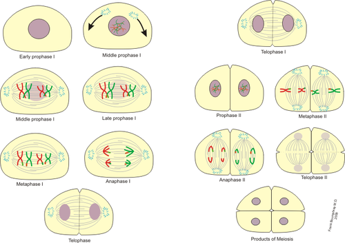
Meiosis
Meiosis (maturing division) is process of reduction division, which takes place in two in a row divisions. The result are cells with haploid number of chromosomes. Germ cells (gamets) arise by meiotic division. The meaning of meiosis rests in a random distribution of paternal and maternal chromosomes into germ cells. This allows for genetic variability. It is increased by crossing-over.
Two different divisions:
- Meiosis I-reduce division(heterotypic). The number of chromosomes is reduced by half. Two haploid cells arise from diploid parent cell.
- Meiosis II- division equation(hemotypic). Sister chromatids are divided into two daughter´s cells.
Meiosis I[edit | edit source]
The reductional division of germ cells, like the division of somatic cells (mitosis), can be divided into four phases:
Prophase I[edit | edit source]
According to the condensation and mutual behavior of duplicated homologous chromosomes, we distinguish a total of 5 stages in prophase I:
Leptotene[edit | edit source]
It is characterized by a low degree of condensation of chromosomes attached at both ends to the inner side of the nuclear envelope (attachment plate). Sister chromatids are close together.
Zygotene[edit | edit source]
Synapses of homologous chromosomes are located at the point of their close attachment. A so-called synaptonemal complex is formed, which connects the two homologues in a zip-like manner. The corresponding loci are always paired next to each other. At the end, all homologues – bivalents (23 in humans) are paired.
- homologous chromosomes find each other by an as yet unknown mechanism
- they begin to pair and gradually adhere to each other along their entire length, the close connection of homologous chromosomes is called a synapse
- the corresponding sections of DNA are usually attached to each other in a linear fashion, creating a structure called a bivalent
- the exception is the X and Y chromosomes in humans, which do not represent a classical homologous pair, but synapse occurs between them
- in the early phase, homologous segments are paired in the so-called pseudoautosomal regions, which occur at both ends of the X and Y chromosomes
- at a later stage, certain adjacent regions are also paired, but most of the length of X and Y remains unpaired
- the synapse formation process is also more complicated in the case of chromosomal structural aberrations, when structures of different shapes are created when homologous regions are paired
- in the presence of inversion, the synapse causes the formation of an inversion loop, in the case of reciprocal translocation, a tetravalent is formed, in the case of Robertsonian translocation, a trivalent, etc.)
Pachytene[edit | edit source]
It begins with the completion of bivalents - sister chromatids are visible as so-called tetrads. Recombination junctions (chiasmata) are the crossing points of non-sister chromatids. Subsequently, crossing-overs occur - recombination of parts of homologues.
- the homologous pairing process is completed
- chromosomes tightly connected using a special protein structure – the synaptonemal complex
- the complex consists of two lateral elements and a central ladder-like element in the middle
- each lateral complex represents the protein axis of one homologous chromosome and is longitudinally attached to both sister chromatids
- loops of chromatin fibers extend radially from the lateral element, and the central element is connected to the lateral ones by transverse filaments
- they remain in this state for several days
- the basic purpose of the synaptonemal complex is to ensure the exact pairing of the corresponding stretches of DNA, thereby contributing to error-free recombination
- the spiraling of the chromosomes continues, they now have the appearance of coarser fibers with unevenly staining areas
- the two-chromatid composition of individual chromosomes begins to be observed
- DNA recombination between non-sister chromatids of homologous chromosomes also takes place at this stage - crossing over (crossing over and exchange of chromosomal segments)
- the actual exchange of DNA segments can also occur between sister chromatids, but in this case it is not recombination in the true sense of the word, since the exchanged segments carry identical genetic makeup
- the probable place where the crossing over process takes place corresponds to the place of occurrence of the so-called recombination nodule (nodule)
Diplotene[edit | edit source]
It is initiated by desynapsis of homologous chromosomes. Disruption of synaptonemal complexes occurs in it. Bivalents remain connected in one or more places (chiasmata).
- homologous chromosomes begin to separate, the synaptonemal complex breaks down (desynapse)
- in the places of crossing over, the chromosomes remain connected to each other, these places are so-called chiasmata
- the distribution of chiasmata is not regular, they can occur anywhere on the chromosome
- however, there are places where crossing over occurs more often - recombination "hotspots" and, conversely, places with a low probability of occurrence (e.g. pericentromeric region)
- it is also possible to observe the phenomenon of chiasmat interference, i.e. reducing the likelihood of a chiasm forming near the appearance of another (the mechanism is still unclear)
- in humans we usually find 1-3 chiasmata on each homologous pair (according to the size of the chromosomes), in total we find 40-50 chiasmata in one cell
- to ensure correct segregation in the further course of the first meiotic division, at least one chiasma occurs even on the smallest chromosome
- four chromatids are already clearly recognizable in each bivalent, the entire structure of a homologous pair of chromosomes is called a tetrad
Diakinesis[edit | edit source]
Diakinesis is the transition to metaphase. There is a strong condensation of chromatids, their division and the release of their end parts from the nuclear envelope. Sister chromatids are joined at distant centromeres of both original homologous chromosomes. The nuclear envelope is disintegrating.
Metaphase I[edit | edit source]
- separate centromeres and kinetochores
- sister chromatids form dyads – by separation of homologous chromosomes
- at the beginning, the nuclear envelope disappears, as in mitosis
- a dividing spindle is formed
- pairs of homologous chromosomes are attached to the microtubules of the dividing apparatus by means of kinetochores and line up in the equatorial plane of the cell
- chiasmata keep homologous chromosomes together until anaphase I begins
- first the chiasmata are located at the point of crossing over, later they move towards the ends of the chromatids (terminalization of the chiasmata)
Anaphase I[edit | edit source]
- homologous chromosomes separate – principle of reduction division
- after the separation of the chiasmata, the homologous chromosomes diverge to opposite poles of the spindle, each consisting of two sister chromatids
- this moment is key for the entire meiosis – the diploid number is reduced to the haploid number
- the distribution of chromosomes of maternal and paternal origin to the poles occurs completely randomly and independently, according to Mendel's law of independent combinability of traits
- a large number of different combinations are created, in humans 233, and with the participation of crossing over the number of combinations is even greater
Telophase I[edit | edit source]
- two cells with different representations of maternal and paternal chromosomes are separated
- haploid sets of chromosomes are clustered at the poles of the cell, two daughter nuclei are formed and, after the division of the cytoplasm, two daughter cells
- this phase is short, the course is very variable
- followed by interkinesis (analogy to mitotic interphase, but DNA replication does not occur)
- chromosomes are temporarily despiralized, interkinesis is very short and in some species completely absent
- the cell then enters II. matures division
Meiosis II[edit | edit source]
It begins after a short interphase during which there is no DNA replication, only decondensation and synthesis of RNA and histones. Subsequent division is a process very similar to mitosis. In prophase II, the centrioles separate and a mitotic spindle is formed, in metaphase II the centromeres and kinetochores of the sister chromatids are oriented to the opposite poles of the dividing spindle. In anaphase II, chromatids separate into daughter cells.
Links[edit | edit source]
Related articles[edit | edit source]
External links[edit | edit source]
Bibliography[edit | edit source]
- ALBERTS,, et al. Základy buněčné biologie. 2.edice edition. 2007. ISBN 80-906902-2-0.
- ŠTEFÁNEK, Jiří. Medicína, nemoci, studium na 1. LF UK [online]. [cit. 11. 2. 2010]. <https://www.stefajir.cz/>.


