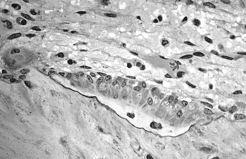
Microscopic structure of bone tissue
Bone is one of the hardest tissues in the human body. It consists of cells and intercellular mass (formed by these cells, it is predominant in volume). The intercellular mass contains a fibrous and amorphous component . Bone is mineralized by calcium salts → difficult processing of bone for histological examination:
- scraping of bone tissue (embedded in Canadian balsam) – cells are not preserved, the bone matrix can be studied;
- common histological methods for decalcification (Bouin's fluid, embedding in celodal or celloidin) - cells remain preserved.
The surface of bone tissue[edit | edit source]
Lined on the surface with layers of collagen fibers → endosteum on the inner surface and periosteum on the outer surface. Both endosteum and periosteum have an outer fibrous layer and an inner layer - osteoblast precursors (osteoprogenitor cells, preosteoblasts) are stored here. The endosteum is thinner than the periosteum, consisting of a thin inner layer of flattened preosteoblasts and a small amount of fibrous tissue (here, numerous small vessels). The outer layer of the periosteum consists of dense collagenous tissue. We also find fibroblasts, collagen fibers, blood vessels and nerve fibers here.
- Sharpey fibers – bundles of collagen fibers penetrating from the outer layer of the periosteum into the bone matrix, firmly connect the periosteum to the bone.
The inner layer of the periosteum (cambium) contains preosteoblasts. These are direct precursors of osteoblasts (osteoprogenitor cells). They are flat, without the ability to divide. They have the character of undifferentiated cells. Outside the preosteoblasts is a small Golgi apparatus and a poorly developed rough endoplasmic reticulum. In the resting phase, these cells do not show high biosynthetic activity, but they can be activated and differentiate into osteoblasts. They play an important role in the process of bone growth and healing (during healing, they can differentiate not only into osteoblasts, but also into chondroblasts or fibroblasts). Their main function is primarily the nutrition of bone tissue, a source of new osteoblasts for the growth, remodeling and replacement of bone tissue (less than periosteum).
Bone tissue cells[edit | edit source]
Bone tissue cells include osteoblasts, osteocytes (precursors – preosteoblasts ) and osteoclasts.
Osteoblasts[edit | edit source]
They lie next to each other on the surface of the beams of bone tissue. The arrangement is similar to the arrangement of cells in a single-layered epithelium. Osteoblasts send out processes that touch each other and lengthen as the cells begin to surround the newly synthesized matrix. They have the character of cells synthesizing proteins for export. If they are actively synthesizing matrix, they are cubic (low cylindrical), have basophilic cytoplasm, and show high alkaline phosphatase activity . With a decrease in synthetic activity, production of alkaline phosphatase flattens and decreases. They have a large bright nucleus, a massive GER, a large GK (under the nucleus), numerous secretory vesicles in the basal area of the cells. Synthesized substances are excreted from the cell on the surface that is in contact with the bone matrix. Osteoblasts produce the organic components of the bone matrix – collagen I, glycosaminoglycans, proteoglycans, glycoproteins. Osteoid is a newly synthesized matrix near osteoblasts that has not yet been mineralized. The deposition of inorganic substances in the bone matrix is also dependent on the presence of osteoblasts. Osteoblasts do not divide, once surrounded by intercellular mass, they become osteocytes (under certain conditions, osteocytes can dedifferentiate back into osteoblasts or preosteoblasts).
Osteocytes[edit | edit source]
Osteocytes are stored individually in lacunae. They send out thin, long processes of cytoplasm that are stored in narrow canals in the bone matrix ( canaliculi ossium ). The processes of neighboring osteocytes are in contact with numerous nexuses . Using nexus, osteocytes communicate with each other, but also with the inner and outer surface of the bone (nutrient supply). Sometimes this chain can form up to 15 cells. Compared to osteoblasts, osteocytes have a smaller nucleus with more heterochromatin. They also have less developed GER, smaller GK and a small number of lysosomes. After resorption, the bone tissues degenerate or turn into osteoblasts again. They are necessary for the existence of the intercellular matrix, they have the ability to synthesize the matrix to a small extent and also participate in its resorption.
Osteoclasts[edit | edit source]
These are free cells of bone tissue. Osteoclasts are large motile cells with numerous processes. It is located on the surface of bone tissue in small depressions → Howship's lacunae. They contain 2–50 nuclei, a large amount of acidophilic cytoplasm with numerous free polysomes. They have not very developed GER, GK, numerous mitochondria and a lot of lysosomes. Osteoclasts have a complex structure of the osteoclast surface - they create so-called invaginations, which significantly increase the resorption surface of the cell → wavy edge. Separated from the rest of the cytoplasm by a light zone without cell organelles (only numerous elements of the cytoskeleton). Lysosomes empty into the area below the wavy edge, creating a low pH environment (the presence of organic acids that osteoclasts produce to a small extent). In the folds of the wavy edge there are numerous crystals of calcium salts and small vesicles.
Osteoclasts belong to the monocytomacrophage system . They develop from a hemocytoblast (a hemopoietic stem cell in the bone marrow ). The immediate precursors of osteoclasts are monocytes (osteoclasts are formed by their fusion).
Intercellular mass[edit | edit source]
Intercellular matter has two components - fibrous and amorphous.
- Fibrous component – collagen fibers (collagen I) – 95% of the organic matter of bone.
- Amorphous component – proteoglycans containing chondroitin sulfate and keratan sulfate, structural glycoproteins (isolated osteonectin, sialoprotein, osteocalcin).
Inorganic substances make up 50% of the dry weight of the bone matrix.
- Calcium ions, phosphate ions, magnesium, potassium, sodium ions, citrate and carbonate ions.
- Calcium and phosphate ions mainly in the form of hydroxyapatite crystals (dimensions 40 × 25 × 3 nm).
- On the surface of the crystals, hydroxyapatite molecules are found in a hydrated form – a layer of saturated solution facilitates the release of calcium ions from the bones.
- The connection of collagen fibers and hydroxyapatite is the reason for the strength of bone tissue.
Links[edit | edit source]
Related articles[edit | edit source]
Sources[edit | edit source]
- JUNQUEIRA, L. Carlos – CARNEIRO, José – KELLEY, Robert O.. Základy histologie. 7. edition. H & H, 1997. pp. 502. ISBN 80-85787-37-7.


