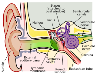
Middle ear
The middle ear is located within the temporal bone extending from the tympanic membrane to the lateral wall of the inner ear. The main function of the middle ear is to transmit vibrations from the tympanic membrane to the inner ear via the auditory ossicles.
Structure[edit | edit source]
The middle ear can be divided into two parts:
- Tympanic cavity – contains the auditory ossicles, which consists of the three small bones known as the malleus, incus and stapes. They transmit sound vibrations through the middle ear.
- Epitympanic recess –which lies next to the mastoid air cells( which releases air into the tympanic cavity when the pressure is low) above the tympanic cavity. The malleus and incus extend upwards slightly entering the epitympanic recess.
Tympanic cavity[edit | edit source]
The tympanic cavity is located between the external ear and the internal ear. This narrow, irregular space has a vertical diameter of about 18 mm, an anteroposterior diameter of about 10 mm, and a transverse diameter of 3 (5) mm. The chorda tympani, a branch of the facial nerve, traverses the tympanic cavity.Its epithelium is single-layered, flattened to cuboid.[edit | edit source]
The tympanic cavity has six boundaries[edit | edit source]
The lateral (or membranous) wall formed by the tympanic membrane and, to a lesser extent, of bone (the squama above, and the tympanic element below). The medial (or labyrinthine) wall constitutes the lateral wall of the internal ear with a promontary in the center formed by the cochlea's basal turn. Fenestra cochleae or the round window lies posteriorly, superiorly lies the oval window or fenestra vestibuli, which is attached to the base of the stapes. Through these windows infection and inflammation spreads to the labyrinth. At the end of the bony canal of the tensor tympani muscle, there is a process around which the tendon of tensor tympani bends. The tympanic plexus innervates the parotid salivary gland that lies under the mucous membrane of the promontory. The roof (or tegmental wall) is formed by Tegmen tympani, petrous part of the temporal bone, and its upper surface is at the floor of the middle cranial fossa. The posterior (or mastoid) wall is directed towards the mastoid process, superiorly opens into the mastoid antrum leading to the mastoid air cells. On the medial wall of its entrance to the antrum has the facial nerve canal and the eminence of the lateral semicircular canal. Below it the stapedius muscle lies in a bony pyramidal eminence. The floor (or jugular wall) has thin plate of bone that lies immediately above the superior bulb of the internal jugular vein. The anterior (or carotid) wall is formed inferiorly by the wall of the internal carotid artery canal. The carotico-tympanic canaliculi for nerves and vessels communicates with the tympanic cavity. The tympanic orifice of the auditory tube lies superiorly.
Nerve and Blood supply of the tympanic cavity[edit | edit source]
The tympanic plexus from the facial nerve (CN VII), the glossopharyngeal nerve(CN IX), and the internal carotid plexus. The anterior tympanic artery (a branch of the maxillary artery), the superior tympanic artery (a branch of the middle meningeal artery), the posterior tympanic artery (a branch of the stylomastoid artery) travels along with the chorda tympani and the inferior tympanic artery (a branch of the pharyngeal artery). Tympanic veins drains into the pterygoid plexus and pharyngeal plexus.[edit | edit source]
Lymph vessels - Regional lymph nodes embedded in the parotid salivary gland.
Auditory ossicles[edit | edit source]
Consists of three bones which are linking the tympanic membrane to the oval window of the internal ear.
- The malleus - largest and most lateral of the ear bones. The handle of the malleus attaches to the tympanic membrane. The head of the malleus articulates with the body of the incus
- The incus – consists of a body and two limbs. The long limb joins the stapes.
- The stapes is the smallest bone in the human body. It joins the incus to the oval window of the inner ear. It contains a head, two limbs, and a base. The head articulates with the incus, and the base joins the oval window.
Auditory tube[edit | edit source]
The auditory tube (eustachian tube) is a cartilaginous and bony tube that connects the middle ear to the nasopharynx. It aims to equalise the middle ear pressure and the pressure of the external auditory meatus.However the pathway can increase the risk of upper respiratory infections that can spread into the middle ear especially the tube is shorter ,straighter and more horizontal in children, therefore middle ear infections tend to be more common in children than adults.
Clinical Significance[edit | edit source]
Otitis Media/ Glue ear and mastoiditis/ mastoid abscess with risk of spreading to middle cranial fossa forming life threatening brain abscess.
Reference[edit | edit source]
The organ of hearing and balance. Torsten Liem DO Osteopath GOsC (GB), in Cranial Osteopathy (Second Edition), 2004.
The Middle Ear - Parts - Bones - Muscles - TeachMeAnatomy. Accessed on 15/04/2024.

