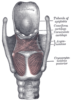
Muscles of the larynx
The larynx is the part of the respiratory system that connects the pharynx to the trachea. Its wall is mainly made up of several cartilages (mainly the unpaired cartillago thyroidea, c. cricoidea and epiglottis, the paired c. arytenoidea, on which the small c. corniculata mounts, further in the plica arytenoidea the c. cuneiformis forms the bump of the tuberculum cuneiforme). The mutual positions of these cartilages determine the shape and width of the glottis (rima glottidis), the tension of the vocal cords, and thus participate in phonation and voice production (in the phonation position). In contrast, in the respiratory position, they keep the glottis wide open and allow free passage of air.
Muscle groups and innervations[edit | edit source]
The seven pairs of laryngeal muscles are divided into 3 groups: ventral (1 muscle), lateral and dorsal (3 muscles each). Innervation comes from branches of the vagus nerve. For the muscle of the ventral group, it is the ramus externus of the superior laryngeal nerve. For the other muscles of the lateral and dorsal groups, it is the recurrent laryngeal nerve.
Ventral group[edit | edit source]
Cricothyroid muscle
- Beginning: outer surface of the arc of the annular cartilage
- Insertion: the lower edge of the thyroid cartilage plate
- Innervation: Nervus laryngeus superior, ramus externus
- Function: tightens and lengthens the vocal folds
It connects to the constrictor pharyngis inferior muscle and together with it forms a ring surrounding the pharynx and larynx. From its beginning, it spreads in a fan-like fashion towards the attachment. The medial bundles are designated as pars recta , the laterally located oblique bundles form pars obliqua.
Lateral group[edit | edit source]
Lateral cricoarytenoid muscle
- Beginning: upper edge of the arc of the annular cartilage
- Attachment: processus muscularis of the vocal cartilage
- Innervation: Nervus laryngeus recurrentens
- Function: adduction of the intermembranous part of the vocal cords
Thyroarytenoid muscle
- Beginning: inner surface of the thyroid cartilage plate
- Insertion: anterolateral surface of the vocal cartilage
- Innervation: Nervus laryngeus recurrentens
- Function: shortens the vocal folds
Its medial part pressing on the ligamentum vocalis inside the plica vocalis is called the musculus vocalis. This changes the thickness of the vocal folds and ensures fine tuning of the glottis.
Thyroepiglottic muscle
- Beginning: inner surface of the thyroid cartilage plate
- Insertion: edge of the epiglottis
- Innervation: Nervus laryngeus recurrentens
- Function: tilts the epiglottis, pulls it towards the root of the tongue and thus widens the entrance to the larynx
It is the cranial continuation of the thyroarytenoid muscle.
Dorsal group[edit | edit source]
Musculus cricoarytenoideus posterior (posticus)
- Beginning: triangular box on the posterior surface of the annular cartilage
- Attachment: processus muscularis of the vocal cartilage
- Innervation: Nervus laryngeus recurrentens
- Function: abduction of vocal cords ( !!! )
As the only muscle of the larynx, it opens the glottis and keeps it open during breathing in the respiratory position. There is a risk of suffocation if its innervation is disturbed (a risk during thyroid operations). If innervation cannot be restored, laterofixation or vocal cord resection should be performed. Its bundles converge towards the insertion.
Arytenoid muscle
- Beginning: vocal cords (see below)
- Attachment: vocal cartilages (see below)
- Innervation: Nervus laryngeus recurrentens
- Function: closes the intercartilaginous part of the glottis
It lies on the dorsal surface of the vocal cords and between them. It passes from one side to the other without an inset tendon inscription. Oblique, more superficial bundles are called pars obliqua, less numerous and deeper bundles form pars transversa.
The aryepiglottic muscle
- Beginning: vocal cartilage
- Attachment: laterocranial edge of the epiglottis
- Innervation: Nervus laryngeus recurrentens
- Function: lowers the epiglottis
It is a continuation of the arytenoideus obliquus muscle (pars obliqua previously muscle, arytenoideus muscle).
Links[edit | edit source]
Related Articles[edit | edit source]
Externí odkazy[edit | edit source]
References[edit | edit source]
- GRIM, Miloš – DRUGA, Rastislav, et al. Základy anatomie 3 : Trávicí, dýchací, močopohlavní a endokrinní systém. 1. edition. Prague : Galén, 2005. 163 pp. ISBN 80-7262-302-8.


