Neuromuscular junction
The neuromuscular (or myoneural) junction is a special type of chemical synapse . Its function is the transmission of excitation from the neuron to the skeletal muscle fiber.
Structure[edit | edit source]
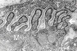
- The presynaptic formation is represented by the axonal end of the motoneuron and is deposited in shallow grooves formed by the invagination of the sarcolemma.
- The postsynaptic structure is represented by the sarcolemma (the plasma membrane of a skeletal muscle fiber).
- The primary synaptic cleft is the space between the presynaptic terminal and the muscle fiber.
- The secondary synaptic cleft is the space created by the secondary invagination of the sarcolemma; the purpose of these invaginations is to increase the receptive surface of the synapse.
- The sarcolemma is lined by a basement membrane, which is why the synaptic cleft at the neuromuscular junction is wider (50−70 nm) than at interneuronal synapses.
The connection itself creates the terminal branches of axons (telodendria), which lose their myelin sheath towards the end, with the sarcolemma of muscle fibers. These endings contain an abundance of small, clear vesicles with acetylcholine, which mediates these connections.
Transmission of excitation to neuromuscular junctions[edit | edit source]
An impulse arriving at a nerve terminal causes exocytosis of synaptic vesicles and release of the mediator into the synaptic cleft. The mediator in the neuromuscular plate is acetylcholine (ACh - it is synthesized in the nerve endings from choline and acetyl coenzyme A). An impulse that reaches the motor neuron ending (telodendria) by depolarization opens the calcium channel and releases about 7000 acetylcholine molecules from the vesicles stored in the terminal part of the nerve. By releasing acetylcholine by exocytosis, a signal for the formation of AP on the sarcolemma is transmitted through nicotinic receptors . Activation of these receptors causes opening of chemically gated Na+ channels and Na+ influx into the cell (based on the concentration gradient) causes a local depolarization (junction potential) that spreads to both sides of the plate. A muscle cell can respond to every stimulus that arrives at the nerve ending with an action potential (this is determined by the size of the disc, the number of activated receptors and the density of voltage-gated Na + channels near the disc). Spontaneous emptying of one vesicle with acetylcholine activates thousands of N-choline receptors, emptying of about 100 vesicles with subsequent opening of about 200,000 channels is necessary to equip a postsynaptic action potential: nerve-induced junction current is formed about 400 nA in size. In order for neuromuscular transmission to function normally, acetylcholine must be inactivated, i.e. split into 2 ineffective components (acetyl and choline) - the disc membrane can thus repolarize and respond to the further release of acetylcholine. This is done by an enzyme, acetylcholinesterase .
Exocytosis of synaptic vesicles from the nerve terminal occurs not only during an action potential, but also individually during accidental contact of the vesicle with the active part of the presynaptic membrane. In these cases, however, only a small amount of acetylcholine enters the synaptic cleft, so that few nicotinic receptors are activated. The resulting depolarization is less than 1 mV (the so-called miniature junction potential) and therefore does not cause the formation of an action potential on the muscle fiber.
Links[edit | edit source]
Related articles[edit | edit source]
Sources[edit | edit source]
- TROJAN, Stanislav, et al. Lékařská fyziologie. 4. edition. Grada, 2003. pp. 771. ISBN 80-247-0512-5.


